

open end, so that the plug is wetted. Take care not to wet the
sides of the straw with sucrose (see Note 14). The column of
sucrose should fill approximately one third of the straw. Using a
separate needle and syringe, inject a small ~0.5 cm column of
100% VSDP leaving at least 1 cm between the sucrose and the
VS solution. Leave prepared straw at room temperature until
required.
2. Place one 50 μL droplet each of 25%, 65%, and 100% VSDP in a
tissue culture dish. Label each droplet.
3. Place liquid nitrogen, to a level equivalent to at least the length
of the straw, into the vessel capable of accommodating the
straw horizontally. Use the thermocouple to determine the
point above the liquid nitrogen at which the temperature is
À140
C and temporarily affix the thermocouple at this level.
4. Pipette a maximum of 5 oocytes (see Note 11) into 25% VSDP
droplet and leave at room temperature for 3–5 min.
5. Transferring as little of the solution as possible (see Note 15),
pipette the oocytes from the 25% VSDP into 65% VSDP
droplet.
6. As quickly as possible, move oocytes into droplet of
100% VSDP.
7. Immediately draw up a small amount of 100% VSDP into a
glass pipette and collect the oocytes. Transfer the oocytes into
the column of 100% VSDP contained within the prepared
straw.
8. Seal the open end of the straw (see Note 2). Hold straw using
holder and, wearing suitable protective equipment, position
the straw horizontally above the liquid nitrogen at which the
temperature is À140
C (see Note 16). Keep the straw in this
position for 3 min and then plunge the straw into liquid
nitrogen.
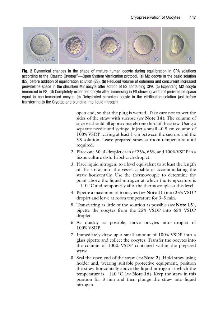
Fig. 2 Dynamical changes in the shape of mature human oocyte during equilibration in CPA solutions
according to the Kitazato Cryotop
®
—Open System vitrification protocol. (a) M2 oocyte in the basic solution
(BS) before addition of equilibration solution (ES). (b) Reduced volume of oolemma and concurrent increased
perivitelline space in the shrunken M2 oocyte after edition of ES containing CPA. (c) Expanding M2 oocyte
immersed in ES. (d) Completely expanded oocyte after immersing in ES showing width of perivitelline space
equal to non-immersed oocyte. (e) Dehydrated shrunken oocyte in the vitrification solution just before
transferring to the Cryotop and plunging into liquid nitrogen
Cryopreservation of Oocytes
447
sides of the straw with sucrose (see Note 14). The column of
sucrose should fill approximately one third of the straw. Using a
separate needle and syringe, inject a small ~0.5 cm column of
100% VSDP leaving at least 1 cm between the sucrose and the
VS solution. Leave prepared straw at room temperature until
required.
2. Place one 50 μL droplet each of 25%, 65%, and 100% VSDP in a
tissue culture dish. Label each droplet.
3. Place liquid nitrogen, to a level equivalent to at least the length
of the straw, into the vessel capable of accommodating the
straw horizontally. Use the thermocouple to determine the
point above the liquid nitrogen at which the temperature is
À140
C and temporarily affix the thermocouple at this level.
4. Pipette a maximum of 5 oocytes (see Note 11) into 25% VSDP
droplet and leave at room temperature for 3–5 min.
5. Transferring as little of the solution as possible (see Note 15),
pipette the oocytes from the 25% VSDP into 65% VSDP
droplet.
6. As quickly as possible, move oocytes into droplet of
100% VSDP.
7. Immediately draw up a small amount of 100% VSDP into a
glass pipette and collect the oocytes. Transfer the oocytes into
the column of 100% VSDP contained within the prepared
straw.
8. Seal the open end of the straw (see Note 2). Hold straw using
holder and, wearing suitable protective equipment, position
the straw horizontally above the liquid nitrogen at which the
temperature is À140
C (see Note 16). Keep the straw in this
position for 3 min and then plunge the straw into liquid
nitrogen.
Fig. 2 Dynamical changes in the shape of mature human oocyte during equilibration in CPA solutions
according to the Kitazato Cryotop
®
—Open System vitrification protocol. (a) M2 oocyte in the basic solution
(BS) before addition of equilibration solution (ES). (b) Reduced volume of oolemma and concurrent increased
perivitelline space in the shrunken M2 oocyte after edition of ES containing CPA. (c) Expanding M2 oocyte
immersed in ES. (d) Completely expanded oocyte after immersing in ES showing width of perivitelline space
equal to non-immersed oocyte. (e) Dehydrated shrunken oocyte in the vitrification solution just before
transferring to the Cryotop and plunging into liquid nitrogen
Cryopreservation of Oocytes
447