

room temperature); (ii) placing an oocyte on the surface of the fine
propylene strip attached to a plastic handle in the minimum
(<1 μL) volume of VS solution (reaching a high cooling rate to
minimise the toxic effect of the CPAs and associated osmotic injury,
also preventing any accidental ice formation, when a lower concentration of CPA is used, and avoiding any fracture of the vitrified
oocyte or surrounding solution); (iii) direct plunging into liquid
nitrogen in an open system thus reaching a high cooling rate
(if required, to avoid contamination of the sample, liquid nitrogen
can be sterilised by filtration or exposure to UV); (iv) rapid warming at 37
C with stepwise equilibration of oocytes in decreasing
concentration of sucrose to prevent the effects of de-vitrification
and osmotic shock resulted in good morphological survival of 95%
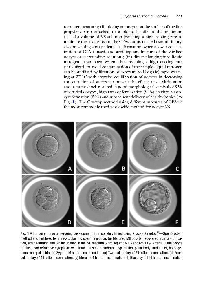
of vitrified oocytes, high rates of fertilization (91%), in vitro blastocyst formation (50%) and subsequent delivery of healthy babies (see
Fig. 1). The Cryotop method using different mixtures of CPAs is
the most commonly used worldwide method for oocyte VS.
Fig. 1 A human embryo undergoing development from oocyte vitrified using Kitazato Cryotop
® —Open System
method and fertilized by intracytoplasmic sperm injection. (a) Matured MII oocyte, recovered from a vitrification, after warming and 3 h incubation in the IVF medium (Vitrolife) at 5% O 2 and 6% CO 2 . After ICSI the oocyte
retains good refractive cytoplasm with intact plasma membrane, typical first polar body, and intact, homogenous zona pellucida. (b) Zygote 16 h after insemination. (c) Two-cell embryo 27 h after insemination. (d) Fourcell embryo 44 h after insemination. (e) Morula 94 h after insemination. (f) Blastocyst 114 h after insemination
Cryopreservation of Oocytes
441
propylene strip attached to a plastic handle in the minimum
(<1 μL) volume of VS solution (reaching a high cooling rate to
minimise the toxic effect of the CPAs and associated osmotic injury,
also preventing any accidental ice formation, when a lower concentration of CPA is used, and avoiding any fracture of the vitrified
oocyte or surrounding solution); (iii) direct plunging into liquid
nitrogen in an open system thus reaching a high cooling rate
(if required, to avoid contamination of the sample, liquid nitrogen
can be sterilised by filtration or exposure to UV); (iv) rapid warming at 37
C with stepwise equilibration of oocytes in decreasing
concentration of sucrose to prevent the effects of de-vitrification
and osmotic shock resulted in good morphological survival of 95%
of vitrified oocytes, high rates of fertilization (91%), in vitro blastocyst formation (50%) and subsequent delivery of healthy babies (see
Fig. 1). The Cryotop method using different mixtures of CPAs is
the most commonly used worldwide method for oocyte VS.
Fig. 1 A human embryo undergoing development from oocyte vitrified using Kitazato Cryotop
® —Open System
method and fertilized by intracytoplasmic sperm injection. (a) Matured MII oocyte, recovered from a vitrification, after warming and 3 h incubation in the IVF medium (Vitrolife) at 5% O 2 and 6% CO 2 . After ICSI the oocyte
retains good refractive cytoplasm with intact plasma membrane, typical first polar body, and intact, homogenous zona pellucida. (b) Zygote 16 h after insemination. (c) Two-cell embryo 27 h after insemination. (d) Fourcell embryo 44 h after insemination. (e) Morula 94 h after insemination. (f) Blastocyst 114 h after insemination
Cryopreservation of Oocytes
441