

2 Materials
2.1
Microwave-Assisted
Drying of Gametes
1. SAM 255 microwave.
2. Karl Fischer titrator (e.g., V20S, Mettler-Toledo,
Columbus, OH).
3. Trehalose dihydrate.
4. Tris/EDTA buffer.
5. α-Hemolysin.
6. Specific materials for gametes (see Table 1).
2.2 Laser-Assisted
Drying of Proteins
1. 1064 nm laser at 5 W.
2. A FLIR SC655 mid-IR camera.
3. Microbalance (e.g., RADWAG).
4. Trehalose dihydrate.
5. Phosphate-buffered saline solution.
6. Saturated solution of lithium chloride (LiCl).
7. Glass coverslips (d ¼ 18 mm).
2.3 Characterization
of Dried Samples
1. Microscope with polarized light imaging.
2. White light interferometer (e.g., a Zygo Nexview scanning
white light interferometer (SWLI) with a 2.75Â Michelson
objective).
3. Raman microscope (e.g., a Horiba LabRAM HR800 confocal
Raman microscope with a 532 nm laser).
3 Methods
Table 2 depicts a comparison of the main parameters used for
microwave- and laser-assisted drying of different types of biologics.
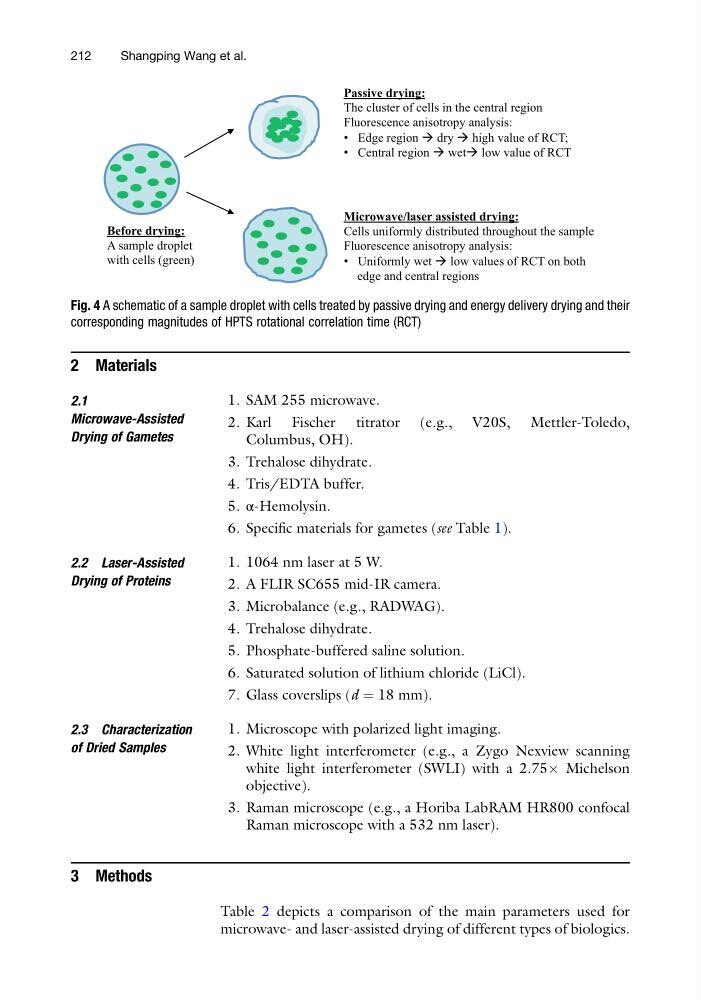
Passive drying:
The cluster of cells in the central region
Fluorescence anisotropy analysis:
• Edge region dry high value of RCT;
• Central region wet low value of RCT
Microwave/laser assisted drying:
Cells uniformly distributed throughout the sample
Fluorescence anisotropy analysis:
• Uniformly wet low values of RCT on both
edge and central regions
Before drying:
A sample droplet
with cells (green)
Fig. 4 A schematic of a sample droplet with cells treated by passive drying and energy delivery drying and their
corresponding magnitudes of HPTS rotational correlation time (RCT)
212
Shangping Wang et al.
2.1
Microwave-Assisted
Drying of Gametes
1. SAM 255 microwave.
2. Karl Fischer titrator (e.g., V20S, Mettler-Toledo,
Columbus, OH).
3. Trehalose dihydrate.
4. Tris/EDTA buffer.
5. α-Hemolysin.
6. Specific materials for gametes (see Table 1).
2.2 Laser-Assisted
Drying of Proteins
1. 1064 nm laser at 5 W.
2. A FLIR SC655 mid-IR camera.
3. Microbalance (e.g., RADWAG).
4. Trehalose dihydrate.
5. Phosphate-buffered saline solution.
6. Saturated solution of lithium chloride (LiCl).
7. Glass coverslips (d ¼ 18 mm).
2.3 Characterization
of Dried Samples
1. Microscope with polarized light imaging.
2. White light interferometer (e.g., a Zygo Nexview scanning
white light interferometer (SWLI) with a 2.75Â Michelson
objective).
3. Raman microscope (e.g., a Horiba LabRAM HR800 confocal
Raman microscope with a 532 nm laser).
3 Methods
Table 2 depicts a comparison of the main parameters used for
microwave- and laser-assisted drying of different types of biologics.
Passive drying:
The cluster of cells in the central region
Fluorescence anisotropy analysis:
• Edge region dry high value of RCT;
• Central region wet low value of RCT
Microwave/laser assisted drying:
Cells uniformly distributed throughout the sample
Fluorescence anisotropy analysis:
• Uniformly wet low values of RCT on both
edge and central regions
Before drying:
A sample droplet
with cells (green)
Fig. 4 A schematic of a sample droplet with cells treated by passive drying and energy delivery drying and their
corresponding magnitudes of HPTS rotational correlation time (RCT)
212
Shangping Wang et al.