

45
Image Filtering, Enhancement, and Restoration
(a)
(b)
(c)
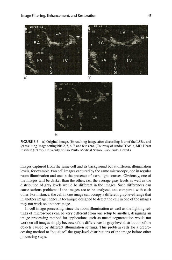
FIGURE 3.6 (a) Original image, (b) resulting image after discarding four of the LSBs, and
(c) resulting image setting bits 2, 5, 6, 7, and 8 to zero. (Courtesy of Andre D’Avila, MD, Heart
Institute (InCor), University of Sao Paulo, Medical School, Sao Paulo, Brazil.)
images captured from the same cell and its background but at different illumination
levels, for example, two cell images captured by the same microscope, one in regular
room illumination and one in the presence of extra light sources. Obviously, one of
the images will be darker than the other; i.e., the average gray levels as well as the
distribution of gray levels would be different in the images. Such differences can
cause serious problems if the images are to be analyzed and compared with each
other. For instance, the cell in one image can occupy a different gray-level range that
in another image; hence, a technique designed to detect the cell in one of the images
may not work on another image.
In cell image processing, since the room illumination as well as the lighting settings of microscopes can be very different from one setup to another, designing an
image processing method for applications such as nuclei segmentation would not
work on all images simply because of the differences in gray-level distribution of the
objects caused by different illumination settings. This problem calls for a preprocessing method to “equalize” the gray-level distributions of the image before other
processing steps.
Image Filtering, Enhancement, and Restoration
(a)
(b)
(c)
FIGURE 3.6 (a) Original image, (b) resulting image after discarding four of the LSBs, and
(c) resulting image setting bits 2, 5, 6, 7, and 8 to zero. (Courtesy of Andre D’Avila, MD, Heart
Institute (InCor), University of Sao Paulo, Medical School, Sao Paulo, Brazil.)
images captured from the same cell and its background but at different illumination
levels, for example, two cell images captured by the same microscope, one in regular
room illumination and one in the presence of extra light sources. Obviously, one of
the images will be darker than the other; i.e., the average gray levels as well as the
distribution of gray levels would be different in the images. Such differences can
cause serious problems if the images are to be analyzed and compared with each
other. For instance, the cell in one image can occupy a different gray-level range that
in another image; hence, a technique designed to detect the cell in one of the images
may not work on another image.
In cell image processing, since the room illumination as well as the lighting settings of microscopes can be very different from one setup to another, designing an
image processing method for applications such as nuclei segmentation would not
work on all images simply because of the differences in gray-level distribution of the
objects caused by different illumination settings. This problem calls for a preprocessing method to “equalize” the gray-level distributions of the image before other
processing steps.