

356
Biomedical Signal and Image Processing
Ocular
Objective
Object
Eye
Image_1=
Object_2
Image_2
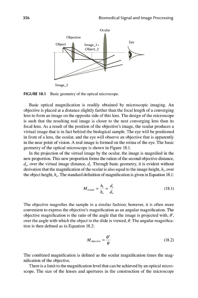
FIGURE 18.1 Basic geometry of the optical microscope.
Basic optical magnification is readily obtained by microscopic imaging. An
objective is placed at a distance slightly farther than the focal length of a converging
lens to form an image on the opposite side of this lens. The design of the microscope
is such that the resulting real image is closer to the next converging lens than its
focal lens. As a result of the position of the objective’s image, the ocular produces a
virtual image that is in fact behind the biological sample. The eye will be positioned
in front of a lens, the ocular, and the eye will observe an objective that is apparently
in the near point of vision. A real image is formed on the retina of the eye. The basic
geometry of the optical microscope is shown in Figure 18.1.
In the projection of the virtual image by the ocular, the image is magnified in the
new proportion. This new proportion forms the ration of the second objective distance,
d o , over the virtual image distance, d i . Through basic geometry, it is evident without
derivation that the magnification of the ocular is also equal to the image height, h i , over
the object height, h o . The standard definition of magnification is given in Equation 18.1:
M ocular =
h i =
d i
(18.1)
h o d o
The objective magnifies the sample in a similar fashion; however, it is often more
convenient to express the objective’s magnification as an angular magnification. The
objective magnification is the ratio of the angle that the image is projected with, θ′,
over the angle with which the object in the slide is viewed, θ. The angular magnification is then defined as in Equation 18.2:
q ′
M objective =
(18.2)
q
The combined magnification is defined as the ocular magnification times the magnification of the objective.
There is a limit to the magnification level that can be achieved by an optical microscope. The size of the lenses and apertures in the construction of the microscope
Biomedical Signal and Image Processing
Ocular
Objective
Object
Eye
Image_1=
Object_2
Image_2
FIGURE 18.1 Basic geometry of the optical microscope.
Basic optical magnification is readily obtained by microscopic imaging. An
objective is placed at a distance slightly farther than the focal length of a converging
lens to form an image on the opposite side of this lens. The design of the microscope
is such that the resulting real image is closer to the next converging lens than its
focal lens. As a result of the position of the objective’s image, the ocular produces a
virtual image that is in fact behind the biological sample. The eye will be positioned
in front of a lens, the ocular, and the eye will observe an objective that is apparently
in the near point of vision. A real image is formed on the retina of the eye. The basic
geometry of the optical microscope is shown in Figure 18.1.
In the projection of the virtual image by the ocular, the image is magnified in the
new proportion. This new proportion forms the ration of the second objective distance,
d o , over the virtual image distance, d i . Through basic geometry, it is evident without
derivation that the magnification of the ocular is also equal to the image height, h i , over
the object height, h o . The standard definition of magnification is given in Equation 18.1:
M ocular =
h i =
d i
(18.1)
h o d o
The objective magnifies the sample in a similar fashion; however, it is often more
convenient to express the objective’s magnification as an angular magnification. The
objective magnification is the ratio of the angle that the image is projected with, θ′,
over the angle with which the object in the slide is viewed, θ. The angular magnification is then defined as in Equation 18.2:
q ′
M objective =
(18.2)
q
The combined magnification is defined as the ocular magnification times the magnification of the objective.
There is a limit to the magnification level that can be achieved by an optical microscope. The size of the lenses and apertures in the construction of the microscope