

346
Biomedical Signal and Image Processing
17.4 PET IMAGE FORMATION
The PET’s tomographic reconstruction algorithm uses the coinciding events measured at all angular and linear positions and reconstructs an image that depicts the
localization and concentration of the positron-emitting radioisotope within a plane
of the organ that was scanned. These sampling features all have an effect on the final
image quality.
For a given pair of gamma rays at angles with a conveniently chosen coordinate
system (θ 1 , θ 2 ) the detector counts n(θ 1 , θ 2 ) coincidence events (see Figure 17.5). The
detector ring has a radius r, which places the location of each annihilation event at
the respective locations with regard to the detectors as rθ 1 and rθ 2 , which are joined
by a line segment l q q
, .
1 2
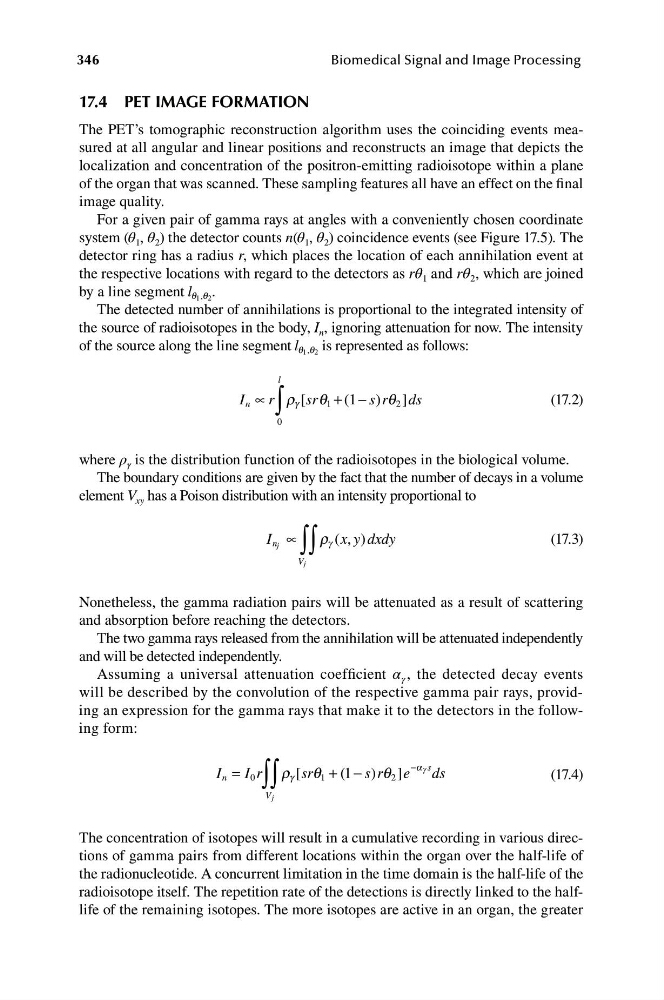
The detected number of annihilations is proportional to the integrated intensity of
the source of radioisotopes in the body, I n , ignoring attenuation for now. The intensity
of the source along the line segment l q q is represented as follows:
,
1 2
l
I n ∝ r g [sr q 1 + (1 − s r q 2 ]ds
∫
r
)
(17.2)
0
where ρ γ is the distribution function of the radioisotopes in the biological volume.
The boundary conditions are given by the fact that the number of decays in a volume
element V xy has a Poison distribution with an intensity proportional to
I j
g x y d x d y
(17.3)
n
( , )
∝
∫∫
r
V j
Nonetheless, the gamma radiation pairs will be attenuated as a result of scattering
and absorption before reaching the detectors.
The two gamma rays released from the annihilation will be attenuated independently
and will be detected independently.
Assuming a universal attenuation coefficient α γ , the detected decay events
will be described by the convolution of the respective gamma pair rays, providing an expression for the gamma rays that make it to the detectors in the following form:
−a g s
I = I r r [sr q + ( 1 − s r
) q ]e ds
n
0
∫∫
g
1
2
(17.4)
V j
The concentration of isotopes will result in a cumulative recording in various directions of gamma pairs from different locations within the organ over the half-life of
the radionucleotide. A concurrent limitation in the time domain is the half-life of the
radioisotope itself. The repetition rate of the detections is directly linked to the halflife of the remaining isotopes. The more isotopes are active in an organ, the greater
Biomedical Signal and Image Processing
17.4 PET IMAGE FORMATION
The PET’s tomographic reconstruction algorithm uses the coinciding events measured at all angular and linear positions and reconstructs an image that depicts the
localization and concentration of the positron-emitting radioisotope within a plane
of the organ that was scanned. These sampling features all have an effect on the final
image quality.
For a given pair of gamma rays at angles with a conveniently chosen coordinate
system (θ 1 , θ 2 ) the detector counts n(θ 1 , θ 2 ) coincidence events (see Figure 17.5). The
detector ring has a radius r, which places the location of each annihilation event at
the respective locations with regard to the detectors as rθ 1 and rθ 2 , which are joined
by a line segment l q q
, .
1 2
The detected number of annihilations is proportional to the integrated intensity of
the source of radioisotopes in the body, I n , ignoring attenuation for now. The intensity
of the source along the line segment l q q is represented as follows:
,
1 2
l
I n ∝ r g [sr q 1 + (1 − s r q 2 ]ds
∫
r
)
(17.2)
0
where ρ γ is the distribution function of the radioisotopes in the biological volume.
The boundary conditions are given by the fact that the number of decays in a volume
element V xy has a Poison distribution with an intensity proportional to
I j
g x y d x d y
(17.3)
n
( , )
∝
∫∫
r
V j
Nonetheless, the gamma radiation pairs will be attenuated as a result of scattering
and absorption before reaching the detectors.
The two gamma rays released from the annihilation will be attenuated independently
and will be detected independently.
Assuming a universal attenuation coefficient α γ , the detected decay events
will be described by the convolution of the respective gamma pair rays, providing an expression for the gamma rays that make it to the detectors in the following form:
−a g s
I = I r r [sr q + ( 1 − s r
) q ]e ds
n
0
∫∫
g
1
2
(17.4)
V j
The concentration of isotopes will result in a cumulative recording in various directions of gamma pairs from different locations within the organ over the half-life of
the radionucleotide. A concurrent limitation in the time domain is the half-life of the
radioisotope itself. The repetition rate of the detections is directly linked to the halflife of the remaining isotopes. The more isotopes are active in an organ, the greater