

336
Biomedical Signal and Image Processing
PROBLEMS
16.1 I ntensity of ultrasound is defined as the pressure per unit of area. An ultrasound wave with a frequency of 5 MHz and an intensity of 20 mW/cm 2 traverses a medium with an acoustic discontinuity at 5 mm depth. Assume there
is no attenuation in either medium. The power collected by the transducer is
0.2 mW over an aperture of 0.15 cm 2 .
a. Calculate the transmitted intensity.
b. Assuming Z = 1.65 × 10 6 kg/m 2
1
s, calculate Z 2 .
c. Calculate the reflected pressure and show that the reflected and transmitted
pressure combined would yield the incident pressure.
16.2 The standard deviation of the collected signal for aortic wall divided by the
mean signal comes close to 1.91 in all four cases. The ratio for muscle tissue
will be less than 1.91. By changing the angle of incidence of the ultrasound
beam and recording two new images at that angle and average them out will
give a new value for each of the tissues that can be used to discriminate
between the lipid, the muscle, the aorta, and aortic wall with plaque. Explain
how adipose tissue (lipids) adjacent to muscle tissue can be distinguished
from aortic wall and from aortic wall with plaque, respectively (Hint: the
solution lies in the scattering properties of the tissues).
16.3 A train of ultrasound pulses is bombarding a slab of medium and is described
by an intensity profile function of time, x(t). The Fourier transform of this periodic phenomenon is X(f). The transmitted ultrasound wave front is y(t) with
Fourier transform, Y(f). The transmitted Fourier transformed wave front is the
characteristic transfer function of the medium, H(f), applied to the incident
Fourier wave front described by the following equation: Y(f) = H(f)X(f). The
transfer function is the Fourier transform of the impulse response function in
the time domain for the tissue slab, h(t). The transmitted ultrasound signal is
collected on the opposite side of the slab after attenuation over the thickness of
the slab.
a. D erive the transfer function when the attenuation coefficient of the
tissue is proportional to the frequency for the frequency range used
in this experiment, α t = α 0 f, where α 0 is the attenuation coefficient at
1 MHz.
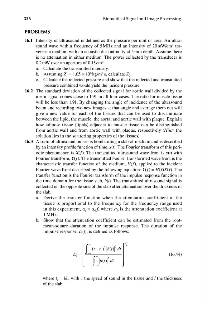
b. S how that the attenuation coefficient can be estimated from the rootmean-square duration of the impulse response. The duration of the
impulse response, D(t), is defined as follows:
⎡
∫
1
∞
2
(
− )
( )
2
⎤
⎢
t t
2
c
h t d
t
⎥
D ⎢
−∞
t =
∞
⎥
(16.44)
⎢
∫
h( )
2
t dt
⎥
⎢ ⎣
−∞
⎦
⎥
where t c = l/c, with c the speed of sound in the tissue and l the thickness
of the slab.
Biomedical Signal and Image Processing
PROBLEMS
16.1 I ntensity of ultrasound is defined as the pressure per unit of area. An ultrasound wave with a frequency of 5 MHz and an intensity of 20 mW/cm 2 traverses a medium with an acoustic discontinuity at 5 mm depth. Assume there
is no attenuation in either medium. The power collected by the transducer is
0.2 mW over an aperture of 0.15 cm 2 .
a. Calculate the transmitted intensity.
b. Assuming Z = 1.65 × 10 6 kg/m 2
1
s, calculate Z 2 .
c. Calculate the reflected pressure and show that the reflected and transmitted
pressure combined would yield the incident pressure.
16.2 The standard deviation of the collected signal for aortic wall divided by the
mean signal comes close to 1.91 in all four cases. The ratio for muscle tissue
will be less than 1.91. By changing the angle of incidence of the ultrasound
beam and recording two new images at that angle and average them out will
give a new value for each of the tissues that can be used to discriminate
between the lipid, the muscle, the aorta, and aortic wall with plaque. Explain
how adipose tissue (lipids) adjacent to muscle tissue can be distinguished
from aortic wall and from aortic wall with plaque, respectively (Hint: the
solution lies in the scattering properties of the tissues).
16.3 A train of ultrasound pulses is bombarding a slab of medium and is described
by an intensity profile function of time, x(t). The Fourier transform of this periodic phenomenon is X(f). The transmitted ultrasound wave front is y(t) with
Fourier transform, Y(f). The transmitted Fourier transformed wave front is the
characteristic transfer function of the medium, H(f), applied to the incident
Fourier wave front described by the following equation: Y(f) = H(f)X(f). The
transfer function is the Fourier transform of the impulse response function in
the time domain for the tissue slab, h(t). The transmitted ultrasound signal is
collected on the opposite side of the slab after attenuation over the thickness of
the slab.
a. D erive the transfer function when the attenuation coefficient of the
tissue is proportional to the frequency for the frequency range used
in this experiment, α t = α 0 f, where α 0 is the attenuation coefficient at
1 MHz.
b. S how that the attenuation coefficient can be estimated from the rootmean-square duration of the impulse response. The duration of the
impulse response, D(t), is defined as follows:
⎡
∫
1
∞
2
(
− )
( )
2
⎤
⎢
t t
2
c
h t d
t
⎥
D ⎢
−∞
t =
∞
⎥
(16.44)
⎢
∫
h( )
2
t dt
⎥
⎢ ⎣
−∞
⎦
⎥
where t c = l/c, with c the speed of sound in the tissue and l the thickness
of the slab.