

300
Biomedical Signal and Image Processing
The BOLD technique relies on the fact that a localized change in blood flow is
observed when the brain neurons are activated and, consequently, the local oxygenation levels change. This increase in oxygenation and hence a decrease of deoxyhemoglobin level results in a change in the MR decay parameter, T 2 * , that can be
measured. These blood flow and oxygenation changes on both vascular and intracellular level have a temporary delay with respect to the neural depolarization sequence
of the cell. This time lag between the physical change in oxygenation level and actual
neural activity is identified as hemodynamic lag.
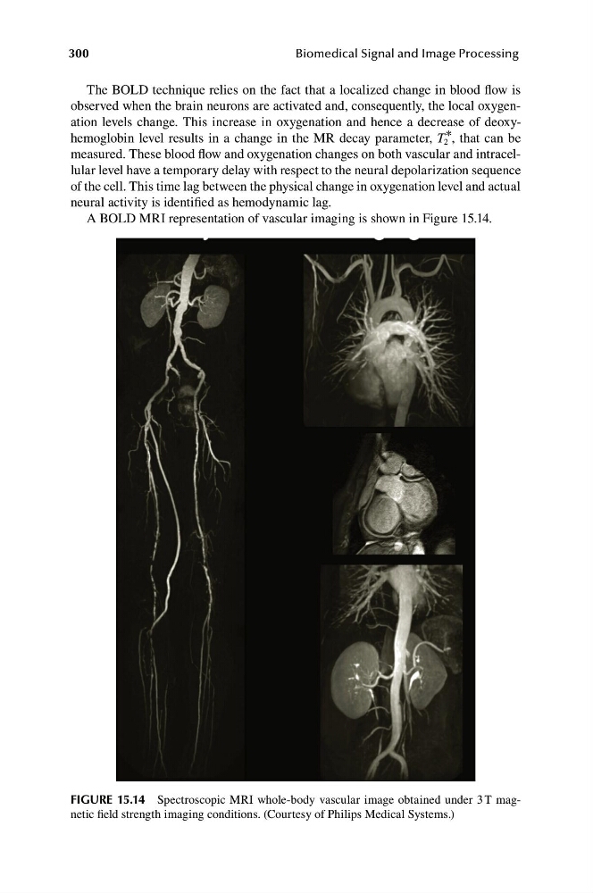
A BOLD MRI representation of vascular imaging is shown in Figure 15.14.
FIGURE 15.14 Spectroscopic MRI whole-body vascular image obtained under 3 T magnetic field strength imaging conditions. (Courtesy of Philips Medical Systems.)
Biomedical Signal and Image Processing
The BOLD technique relies on the fact that a localized change in blood flow is
observed when the brain neurons are activated and, consequently, the local oxygenation levels change. This increase in oxygenation and hence a decrease of deoxyhemoglobin level results in a change in the MR decay parameter, T 2 * , that can be
measured. These blood flow and oxygenation changes on both vascular and intracellular level have a temporary delay with respect to the neural depolarization sequence
of the cell. This time lag between the physical change in oxygenation level and actual
neural activity is identified as hemodynamic lag.
A BOLD MRI representation of vascular imaging is shown in Figure 15.14.
FIGURE 15.14 Spectroscopic MRI whole-body vascular image obtained under 3 T magnetic field strength imaging conditions. (Courtesy of Philips Medical Systems.)