

270
Biomedical Signal and Image Processing
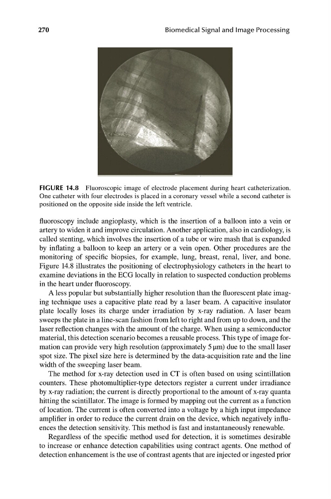
FIGURE 14.8 Fluoroscopic image of electrode placement during heart catheterization.
One catheter with four electrodes is placed in a coronary vessel while a second catheter is
positioned on the opposite side inside the left ventricle.
fluoroscopy include angioplasty, which is the insertion of a balloon into a vein or
artery to widen it and improve circulation. Another application, also in cardiology, is
called stenting, which involves the insertion of a tube or wire mash that is expanded
by inflating a balloon to keep an artery or a vein open. Other procedures are the
monitoring of specific biopsies, for example, lung, breast, renal, liver, and bone.
Figure 14.8 illustrates the positioning of electrophysiology catheters in the heart to
examine deviations in the ECG locally in relation to suspected conduction problems
in the heart under fluoroscopy.
A less popular but substantially higher resolution than the fluorescent plate imaging technique uses a capacitive plate read by a laser beam. A capacitive insulator
plate locally loses its charge under irradiation by x-ray radiation. A laser beam
sweeps the plate in a line-scan fashion from left to right and from up to down, and the
laser reflection changes with the amount of the charge. When using a semiconductor
material, this detection scenario becomes a reusable process. This type of image formation can provide very high resolution (approximately 5 μm) due to the small laser
spot size. The pixel size here is determined by the data-acquisition rate and the line
width of the sweeping laser beam.
The method for x-ray detection used in CT is often based on using scintillation
counters. These photomultiplier-type detectors register a current under irradiance
by x-ray radiation; the current is directly proportional to the amount of x-ray quanta
hitting the scintillator. The image is formed by mapping out the current as a function
of location. The current is often converted into a voltage by a high input impedance
amplifier in order to reduce the current drain on the device, which negatively influences the detection sensitivity. This method is fast and instantaneously renewable.
Regardless of the specific method used for detection, it is sometimes desirable
to increase or enhance detection capabilities using contract agents. One method of
detection enhancement is the use of contrast agents that are injected or ingested prior
Biomedical Signal and Image Processing
FIGURE 14.8 Fluoroscopic image of electrode placement during heart catheterization.
One catheter with four electrodes is placed in a coronary vessel while a second catheter is
positioned on the opposite side inside the left ventricle.
fluoroscopy include angioplasty, which is the insertion of a balloon into a vein or
artery to widen it and improve circulation. Another application, also in cardiology, is
called stenting, which involves the insertion of a tube or wire mash that is expanded
by inflating a balloon to keep an artery or a vein open. Other procedures are the
monitoring of specific biopsies, for example, lung, breast, renal, liver, and bone.
Figure 14.8 illustrates the positioning of electrophysiology catheters in the heart to
examine deviations in the ECG locally in relation to suspected conduction problems
in the heart under fluoroscopy.
A less popular but substantially higher resolution than the fluorescent plate imaging technique uses a capacitive plate read by a laser beam. A capacitive insulator
plate locally loses its charge under irradiation by x-ray radiation. A laser beam
sweeps the plate in a line-scan fashion from left to right and from up to down, and the
laser reflection changes with the amount of the charge. When using a semiconductor
material, this detection scenario becomes a reusable process. This type of image formation can provide very high resolution (approximately 5 μm) due to the small laser
spot size. The pixel size here is determined by the data-acquisition rate and the line
width of the sweeping laser beam.
The method for x-ray detection used in CT is often based on using scintillation
counters. These photomultiplier-type detectors register a current under irradiance
by x-ray radiation; the current is directly proportional to the amount of x-ray quanta
hitting the scintillator. The image is formed by mapping out the current as a function
of location. The current is often converted into a voltage by a high input impedance
amplifier in order to reduce the current drain on the device, which negatively influences the detection sensitivity. This method is fast and instantaneously renewable.
Regardless of the specific method used for detection, it is sometimes desirable
to increase or enhance detection capabilities using contract agents. One method of
detection enhancement is the use of contrast agents that are injected or ingested prior