

229
Electromyogram
kg
μV
0.1 s
9
6
3
0
+20
–20
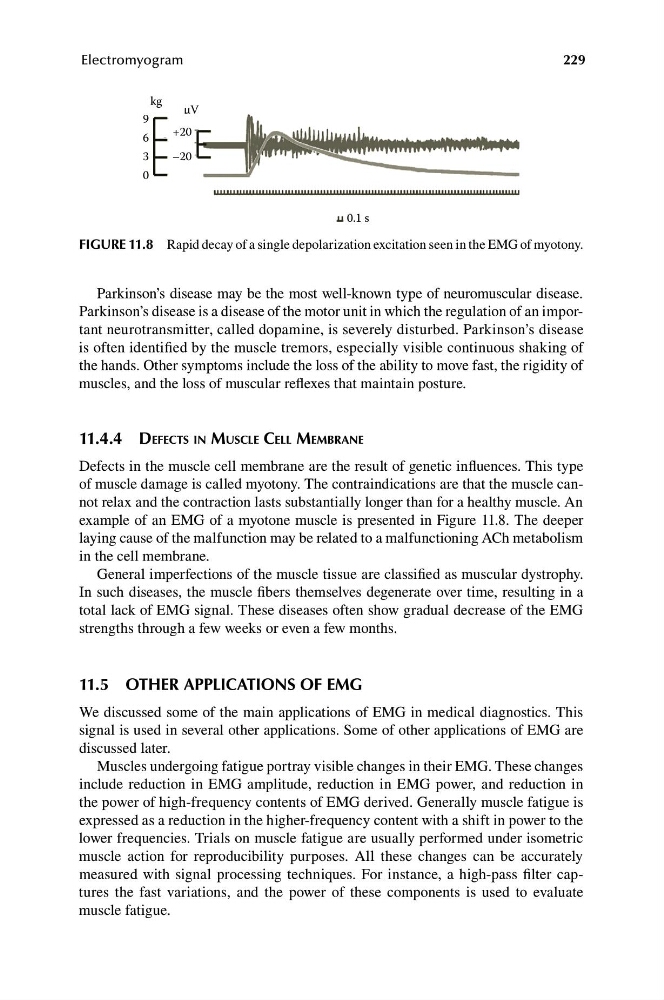
FIGURE 11.8 Rapid decay of a single depolarization excitation seen in the EMG of myotony.
Parkinson’s disease may be the most well-known type of neuromuscular disease.
Parkinson’s disease is a disease of the motor unit in which the regulation of an important neurotransmitter, called dopamine, is severely disturbed. Parkinson’s disease
is often identified by the muscle tremors, especially visible continuous shaking of
the hands. Other symptoms include the loss of the ability to move fast, the rigidity of
muscles, and the loss of muscular reflexes that maintain posture.
11.4.4 DEFECTS IN MUSCLE CELL MEMBRANE
Defects in the muscle cell membrane are the result of genetic influences. This type
of muscle damage is called myotony. The contraindications are that the muscle cannot relax and the contraction lasts substantially longer than for a healthy muscle. An
example of an EMG of a myotone muscle is presented in Figure 11.8. The deeper
laying cause of the malfunction may be related to a malfunctioning ACh metabolism
in the cell membrane.
General imperfections of the muscle tissue are classified as muscular dystrophy.
In such diseases, the muscle fibers themselves degenerate over time, resulting in a
total lack of EMG signal. These diseases often show gradual decrease of the EMG
strengths through a few weeks or even a few months.
11.5 OTHER APPLICATIONS OF EMG
We discussed some of the main applications of EMG in medical diagnostics. This
signal is used in several other applications. Some of other applications of EMG are
discussed later.
Muscles undergoing fatigue portray visible changes in their EMG. These changes
include reduction in EMG amplitude, reduction in EMG power, and reduction in
the power of high-frequency contents of EMG derived. Generally muscle fatigue is
expressed as a reduction in the higher-frequency content with a shift in power to the
lower frequencies. Trials on muscle fatigue are usually performed under isometric
muscle action for reproducibility purposes. All these changes can be accurately
measured with signal processing techniques. For instance, a high-pass filter captures the fast variations, and the power of these components is used to evaluate
muscle fatigue.
Electromyogram
kg
μV
0.1 s
9
6
3
0
+20
–20
FIGURE 11.8 Rapid decay of a single depolarization excitation seen in the EMG of myotony.
Parkinson’s disease may be the most well-known type of neuromuscular disease.
Parkinson’s disease is a disease of the motor unit in which the regulation of an important neurotransmitter, called dopamine, is severely disturbed. Parkinson’s disease
is often identified by the muscle tremors, especially visible continuous shaking of
the hands. Other symptoms include the loss of the ability to move fast, the rigidity of
muscles, and the loss of muscular reflexes that maintain posture.
11.4.4 DEFECTS IN MUSCLE CELL MEMBRANE
Defects in the muscle cell membrane are the result of genetic influences. This type
of muscle damage is called myotony. The contraindications are that the muscle cannot relax and the contraction lasts substantially longer than for a healthy muscle. An
example of an EMG of a myotone muscle is presented in Figure 11.8. The deeper
laying cause of the malfunction may be related to a malfunctioning ACh metabolism
in the cell membrane.
General imperfections of the muscle tissue are classified as muscular dystrophy.
In such diseases, the muscle fibers themselves degenerate over time, resulting in a
total lack of EMG signal. These diseases often show gradual decrease of the EMG
strengths through a few weeks or even a few months.
11.5 OTHER APPLICATIONS OF EMG
We discussed some of the main applications of EMG in medical diagnostics. This
signal is used in several other applications. Some of other applications of EMG are
discussed later.
Muscles undergoing fatigue portray visible changes in their EMG. These changes
include reduction in EMG amplitude, reduction in EMG power, and reduction in
the power of high-frequency contents of EMG derived. Generally muscle fatigue is
expressed as a reduction in the higher-frequency content with a shift in power to the
lower frequencies. Trials on muscle fatigue are usually performed under isometric
muscle action for reproducibility purposes. All these changes can be accurately
measured with signal processing techniques. For instance, a high-pass filter captures the fast variations, and the power of these components is used to evaluate
muscle fatigue.