

xxii
Introduction
Biological
system
Sensors
Preprocessing
and filtering
Feature
extraction
Classification and
diagnostics
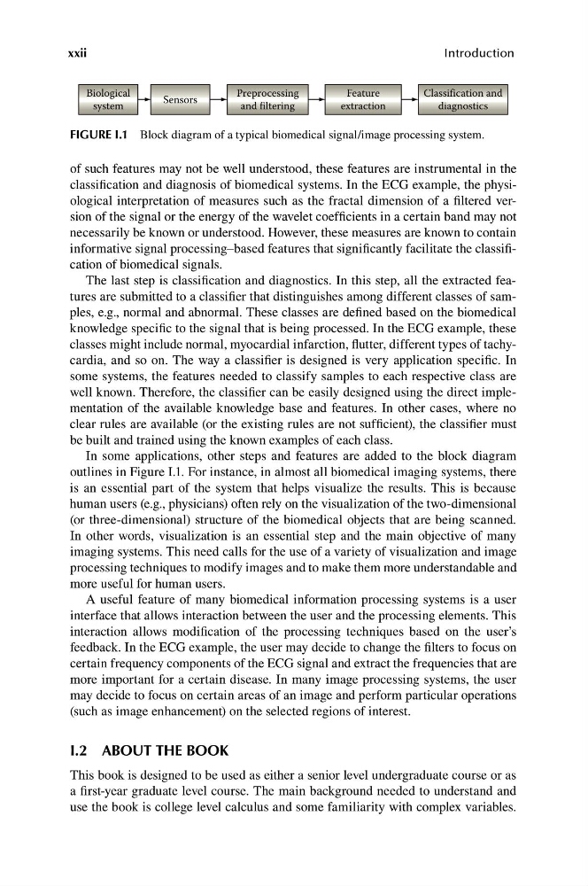
FIGURE I.1 Block diagram of a typical biomedical signal/image processing system.
of such features may not be well understood, these features are instrumental in the
classification and diagnosis of biomedical systems. In the ECG example, the physiological interpretation of measures such as the fractal dimension of a filtered version of the signal or the energy of the wavelet coefficients in a certain band may not
necessarily be known or understood. However, these measures are known to contain
informative signal processing–based features that significantly facilitate the classification of biomedical signals.
The last step is classification and diagnostics. In this step, all the extracted features are submitted to a classifier that distinguishes among different classes of samples, e.g., normal and abnormal. These classes are defined based on the biomedical
knowledge specific to the signal that is being processed. In the ECG example, these
classes might include normal, myocardial infarction, flutter, different types of tachycardia, and so on. The way a classifier is designed is very application specific. In
some systems, the features needed to classify samples to each respective class are
well known. Therefore, the classifier can be easily designed using the direct implementation of the available knowledge base and features. In other cases, where no
clear rules are available (or the existing rules are not sufficient), the classifier must
be built and trained using the known examples of each class.
In some applications, other steps and features are added to the block diagram
outlines in Figure I.1. For instance, in almost all biomedical imaging systems, there
is an essential part of the system that helps visualize the results. This is because
human users (e.g., physicians) often rely on the visualization of the two-dimensional
(or three-dimensional) structure of the biomedical objects that are being scanned.
In other words, visualization is an essential step and the main objective of many
imaging systems. This need calls for the use of a variety of visualization and image
processing techniques to modify images and to make them more understandable and
more useful for human users.
A useful feature of many biomedical information processing systems is a user
interface that allows interaction between the user and the processing elements. This
interaction allows modification of the processing techniques based on the user’s
feedback. In the ECG example, the user may decide to change the filters to focus on
certain frequency components of the ECG signal and extract the frequencies that are
more important for a certain disease. In many image processing systems, the user
may decide to focus on certain areas of an image and perform particular operations
(such as image enhancement) on the selected regions of interest.
I.2 ABOUT THE BOOK
This book is designed to be used as either a senior level undergraduate course or as
a first-year graduate level course. The main background needed to understand and
use the book is college level calculus and some familiarity with complex variables.
Introduction
Biological
system
Sensors
Preprocessing
and filtering
Feature
extraction
Classification and
diagnostics
FIGURE I.1 Block diagram of a typical biomedical signal/image processing system.
of such features may not be well understood, these features are instrumental in the
classification and diagnosis of biomedical systems. In the ECG example, the physiological interpretation of measures such as the fractal dimension of a filtered version of the signal or the energy of the wavelet coefficients in a certain band may not
necessarily be known or understood. However, these measures are known to contain
informative signal processing–based features that significantly facilitate the classification of biomedical signals.
The last step is classification and diagnostics. In this step, all the extracted features are submitted to a classifier that distinguishes among different classes of samples, e.g., normal and abnormal. These classes are defined based on the biomedical
knowledge specific to the signal that is being processed. In the ECG example, these
classes might include normal, myocardial infarction, flutter, different types of tachycardia, and so on. The way a classifier is designed is very application specific. In
some systems, the features needed to classify samples to each respective class are
well known. Therefore, the classifier can be easily designed using the direct implementation of the available knowledge base and features. In other cases, where no
clear rules are available (or the existing rules are not sufficient), the classifier must
be built and trained using the known examples of each class.
In some applications, other steps and features are added to the block diagram
outlines in Figure I.1. For instance, in almost all biomedical imaging systems, there
is an essential part of the system that helps visualize the results. This is because
human users (e.g., physicians) often rely on the visualization of the two-dimensional
(or three-dimensional) structure of the biomedical objects that are being scanned.
In other words, visualization is an essential step and the main objective of many
imaging systems. This need calls for the use of a variety of visualization and image
processing techniques to modify images and to make them more understandable and
more useful for human users.
A useful feature of many biomedical information processing systems is a user
interface that allows interaction between the user and the processing elements. This
interaction allows modification of the processing techniques based on the user’s
feedback. In the ECG example, the user may decide to change the filters to focus on
certain frequency components of the ECG signal and extract the frequencies that are
more important for a certain disease. In many image processing systems, the user
may decide to focus on certain areas of an image and perform particular operations
(such as image enhancement) on the selected regions of interest.
I.2 ABOUT THE BOOK
This book is designed to be used as either a senior level undergraduate course or as
a first-year graduate level course. The main background needed to understand and
use the book is college level calculus and some familiarity with complex variables.