

192
Biomedical Signal and Image Processing
I
aV R
V1
V4
II
aV L
V2
V5
III
(a)
aV F
V3
V6
(b)
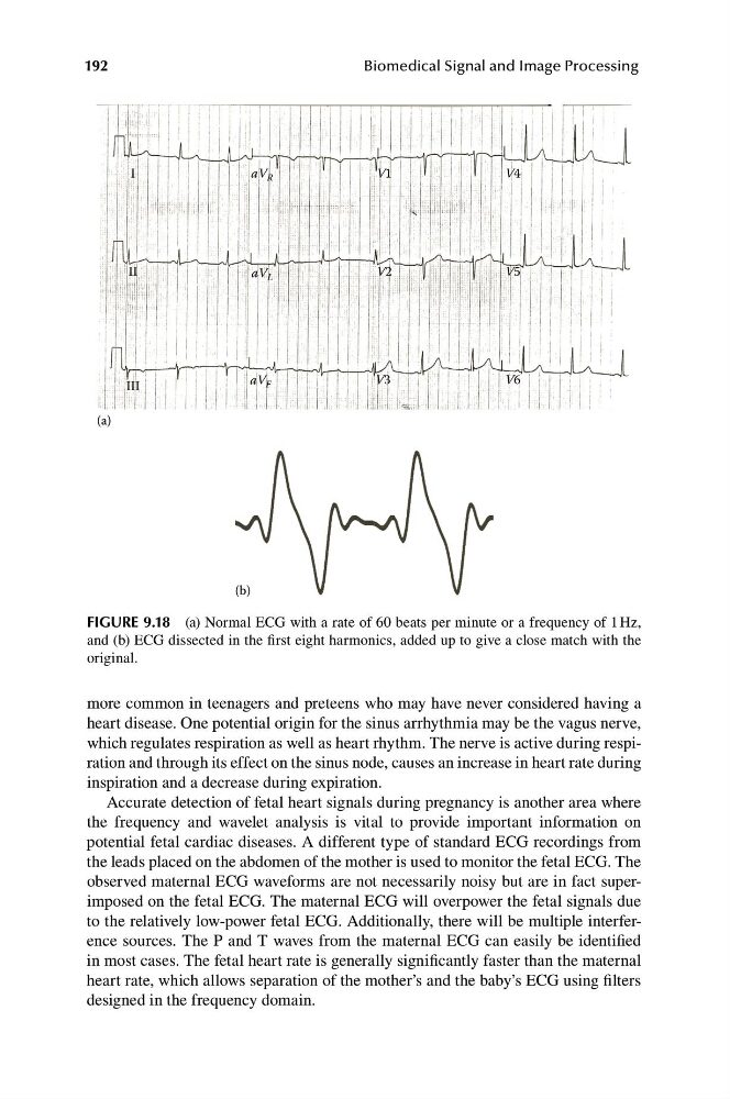
FIGURE 9.18 (a) Normal ECG with a rate of 60 beats per minute or a frequency of 1 Hz,
and (b) ECG dissected in the first eight harmonics, added up to give a close match with the
original.
more common in teenagers and preteens who may have never considered having a
heart disease. One potential origin for the sinus arrhythmia may be the vagus nerve,
which regulates respiration as well as heart rhythm. The nerve is active during respiration and through its effect on the sinus node, causes an increase in heart rate during
inspiration and a decrease during expiration.
Accurate detection of fetal heart signals during pregnancy is another area where
the frequency and wavelet analysis is vital to provide important information on
potential fetal cardiac diseases. A different type of standard ECG recordings from
the leads placed on the abdomen of the mother is used to monitor the fetal ECG. The
observed maternal ECG waveforms are not necessarily noisy but are in fact superimposed on the fetal ECG. The maternal ECG will overpower the fetal signals due
to the relatively low-power fetal ECG. Additionally, there will be multiple interference sources. The P and T waves from the maternal ECG can easily be identified
in most cases. The fetal heart rate is generally significantly faster than the maternal
heart rate, which allows separation of the mother’s and the baby’s ECG using filters
designed in the frequency domain.
Biomedical Signal and Image Processing
I
aV R
V1
V4
II
aV L
V2
V5
III
(a)
aV F
V3
V6
(b)
FIGURE 9.18 (a) Normal ECG with a rate of 60 beats per minute or a frequency of 1 Hz,
and (b) ECG dissected in the first eight harmonics, added up to give a close match with the
original.
more common in teenagers and preteens who may have never considered having a
heart disease. One potential origin for the sinus arrhythmia may be the vagus nerve,
which regulates respiration as well as heart rhythm. The nerve is active during respiration and through its effect on the sinus node, causes an increase in heart rate during
inspiration and a decrease during expiration.
Accurate detection of fetal heart signals during pregnancy is another area where
the frequency and wavelet analysis is vital to provide important information on
potential fetal cardiac diseases. A different type of standard ECG recordings from
the leads placed on the abdomen of the mother is used to monitor the fetal ECG. The
observed maternal ECG waveforms are not necessarily noisy but are in fact superimposed on the fetal ECG. The maternal ECG will overpower the fetal signals due
to the relatively low-power fetal ECG. Additionally, there will be multiple interference sources. The P and T waves from the maternal ECG can easily be identified
in most cases. The fetal heart rate is generally significantly faster than the maternal
heart rate, which allows separation of the mother’s and the baby’s ECG using filters
designed in the frequency domain.