

2.3.2 Merging LBP Features
Performing mean variance normalization on the grayscale image and the corresponding
LBP feature image, and then combining the grayscale image with the LBP image in the
channel dimension, for example, the shape of the grayscale image is (h, w), the shape
of the LBP image Also (h, w), the combined shape is (h, w, 2).
3 The Experiment
3.1 Experimental Dataset
MURA is a dataset of musculoskeletal radiographs, which contains a total of 14,863
studies of 12,173 patients and 40,561 multiview radiographs. Each was one of seven
types of standard upper extremity radiology studies: fingers, elbows, forearms, hands,
humerus, shoulders, and wrists [8].
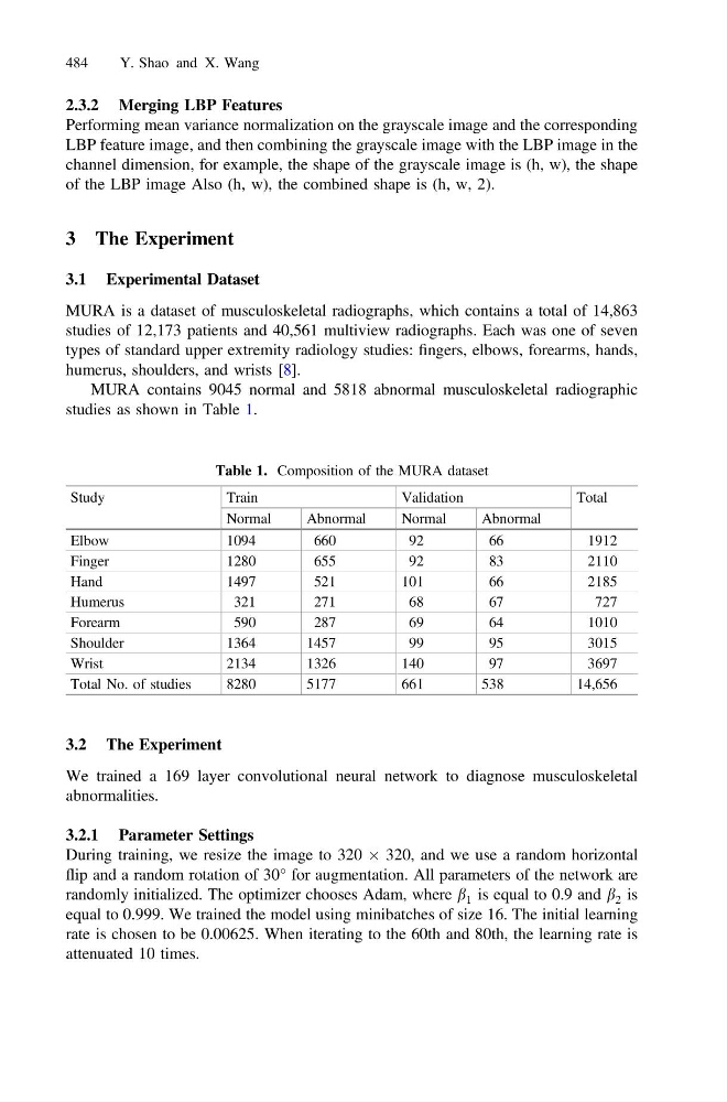
MURA contains 9045 normal and 5818 abnormal musculoskeletal radiographic
studies as shown in Table 1.
3.2 The Experiment
We trained a 169 layer convolutional neural network to diagnose musculoskeletal
abnormalities.
3.2.1 Parameter Settings
During training, we resize the image to 320 Â 320, and we use a random horizontal
flip and a random rotation of 30° for augmentation. All parameters of the network are
randomly initialized. The optimizer chooses Adam, where b 1 is equal to 0.9 and b 2 is
equal to 0.999. We trained the model using minibatches of size 16. The initial learning
rate is chosen to be 0.00625. When iterating to the 60th and 80th, the learning rate is
attenuated 10 times.
Table 1. Composition of the MURA dataset
Study
Train
Validation
Total
Normal
Abnormal
Normal
Abnormal
Elbow
1094
660
92
66
1912
Finger
1280
655
92
83
2110
Hand
1497
521
101
66
2185
Humerus
321
271
68
67
727
Forearm
590
287
69
64
1010
Shoulder
1364
1457
99
95
3015
Wrist
2134
1326
140
97
3697
Total No. of studies
8280
5177
661
538
14,656
484
Y. Shao and X. Wang