

88
4 PVA Bionanocomposite Films with Different Particle …
4.4 XRD Patterns
XRD analysis is a very useful material characterisation method to evaluate crystalline structures of polymers and composites, as well as to determine d-spacing
values between clay interlayers. By monitoring the position and intensity of basal
reflections from distributed silicate layers, nanocomposite structures (i.e. intercalated
or exfoliated), as well as clay aggregated structures can be identified accordingly
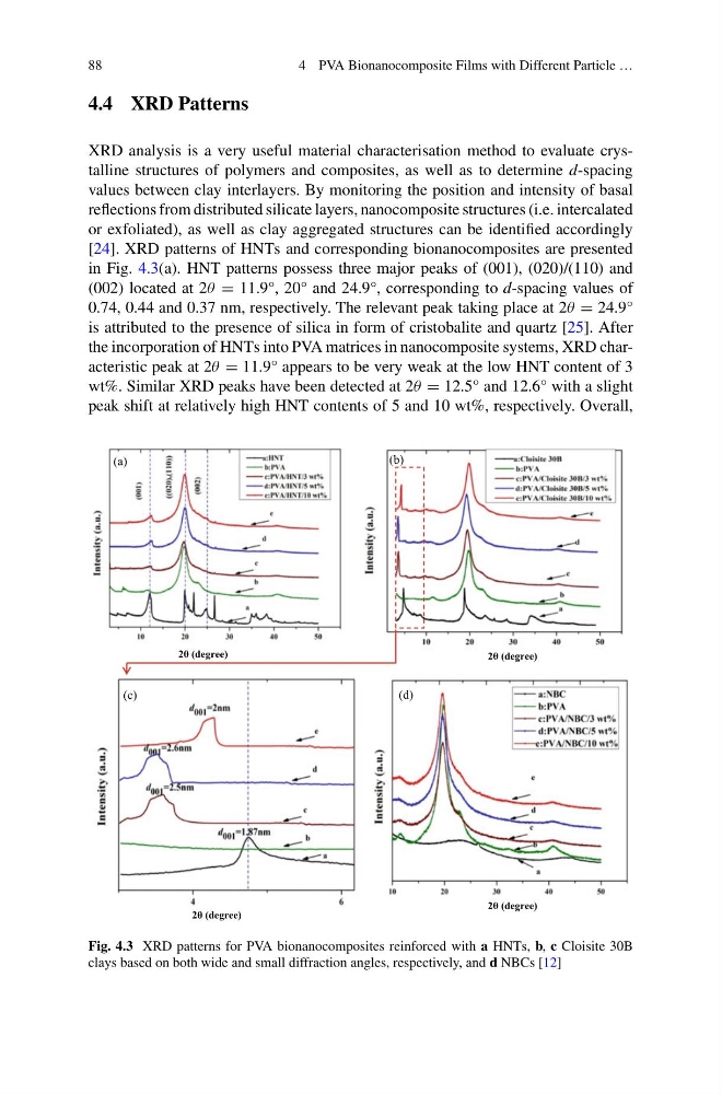
[24]. XRD patterns of HNTs and corresponding bionanocomposites are presented
in Fig. 4.3(a). HNT patterns possess three major peaks of (001), (020)/(110) and
(002) located at 2θ = 11.9°, 20° and 24.9°, corresponding to d-spacing values of
0.74, 0.44 and 0.37 nm, respectively. The relevant peak taking place at 2θ = 24.9°
is attributed to the presence of silica in form of cristobalite and quartz [25]. After
the incorporation of HNTs into PVA matrices in nanocomposite systems, XRD characteristic peak at 2θ = 11.9° appears to be very weak at the low HNT content of 3
wt%. Similar XRD peaks have been detected at 2θ = 12.5° and 12.6° with a slight
peak shift at relatively high HNT contents of 5 and 10 wt%, respectively. Overall,
(b)
(c)
(d)
2θ (degree)
2θ (degree)
2θ (degree)
2θ (degree)
(a)
Fig. 4.3 XRD patterns for PVA bionanocomposites reinforced with a HNTs, b, c Cloisite 30B
clays based on both wide and small diffraction angles, respectively, and d NBCs [12]
4 PVA Bionanocomposite Films with Different Particle …
4.4 XRD Patterns
XRD analysis is a very useful material characterisation method to evaluate crystalline structures of polymers and composites, as well as to determine d-spacing
values between clay interlayers. By monitoring the position and intensity of basal
reflections from distributed silicate layers, nanocomposite structures (i.e. intercalated
or exfoliated), as well as clay aggregated structures can be identified accordingly
[24]. XRD patterns of HNTs and corresponding bionanocomposites are presented
in Fig. 4.3(a). HNT patterns possess three major peaks of (001), (020)/(110) and
(002) located at 2θ = 11.9°, 20° and 24.9°, corresponding to d-spacing values of
0.74, 0.44 and 0.37 nm, respectively. The relevant peak taking place at 2θ = 24.9°
is attributed to the presence of silica in form of cristobalite and quartz [25]. After
the incorporation of HNTs into PVA matrices in nanocomposite systems, XRD characteristic peak at 2θ = 11.9° appears to be very weak at the low HNT content of 3
wt%. Similar XRD peaks have been detected at 2θ = 12.5° and 12.6° with a slight
peak shift at relatively high HNT contents of 5 and 10 wt%, respectively. Overall,
(b)
(c)
(d)
2θ (degree)
2θ (degree)
2θ (degree)
2θ (degree)
(a)
Fig. 4.3 XRD patterns for PVA bionanocomposites reinforced with a HNTs, b, c Cloisite 30B
clays based on both wide and small diffraction angles, respectively, and d NBCs [12]