

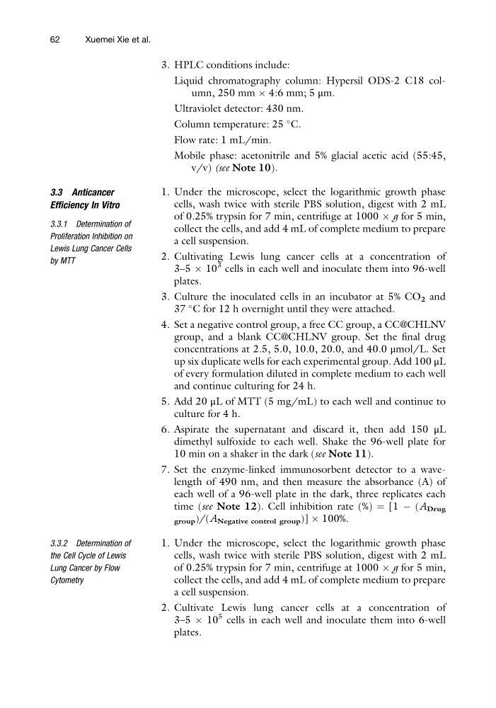
3. HPLC conditions include:
Liquid chromatography column: Hypersil ODS-2 C18 column, 250 mm  4:6 mm; 5 μm.
Ultraviolet detector: 430 nm.
Column temperature: 25
C.
Flow rate: 1 mL/min.
Mobile phase: acetonitrile and 5% glacial acetic acid (55:45,
v/v) (see Note 10).
3.3 Anticancer
Efficiency In Vitro
3.3.1 Determination of
Proliferation Inhibition on
Lewis Lung Cancer Cells
by MTT
1. Under the microscope, select the logarithmic growth phase
cells, wash twice with sterile PBS solution, digest with 2 mL
of 0.25% trypsin for 7 min, centrifuge at 1000 Â g for 5 min,
collect the cells, and add 4 mL of complete medium to prepare
a cell suspension.
2. Cultivating Lewis lung cancer cells at a concentration of
3–5 Â 10
3 cells in each well and inoculate them into 96-well
plates.
3. Culture the inoculated cells in an incubator at 5% CO 2 and
37
C for 12 h overnight until they were attached.
4. Set a negative control group, a free CC group, a CC@CHLNV
group, and a blank CC@CHLNV group. Set the final drug
concentrations at 2.5, 5.0, 10.0, 20.0, and 40.0 μmol/L. Set
up six duplicate wells for each experimental group. Add 100 μL
of every formulation diluted in complete medium to each well
and continue culturing for 24 h.
5. Add 20 μL of MTT (5 mg/mL) to each well and continue to
culture for 4 h.
6. Aspirate the supernatant and discard it, then add 150 μL
dimethyl sulfoxide to each well. Shake the 96-well plate for
10 min on a shaker in the dark (see Note 11).
7. Set the enzyme-linked immunosorbent detector to a wavelength of 490 nm, and then measure the absorbance (A) of
each well of a 96-well plate in the dark, three replicates each
time (see Note 12). Cell inhibition rate (%) ¼ [1 À (A Drug
group )/(A Negative control group )] Â 100%.
3.3.2 Determination of
the Cell Cycle of Lewis
Lung Cancer by Flow
Cytometry
1. Under the microscope, select the logarithmic growth phase
cells, wash twice with sterile PBS solution, digest with 2 mL
of 0.25% trypsin for 7 min, centrifuge at 1000 Â g for 5 min,
collect the cells, and add 4 mL of complete medium to prepare
a cell suspension.
2. Cultivate Lewis lung cancer cells at a concentration of
3–5 Â 10
5 cells in each well and inoculate them into 6-well
plates.
62
Xuemei Xie et al.
Liquid chromatography column: Hypersil ODS-2 C18 column, 250 mm  4:6 mm; 5 μm.
Ultraviolet detector: 430 nm.
Column temperature: 25
C.
Flow rate: 1 mL/min.
Mobile phase: acetonitrile and 5% glacial acetic acid (55:45,
v/v) (see Note 10).
3.3 Anticancer
Efficiency In Vitro
3.3.1 Determination of
Proliferation Inhibition on
Lewis Lung Cancer Cells
by MTT
1. Under the microscope, select the logarithmic growth phase
cells, wash twice with sterile PBS solution, digest with 2 mL
of 0.25% trypsin for 7 min, centrifuge at 1000 Â g for 5 min,
collect the cells, and add 4 mL of complete medium to prepare
a cell suspension.
2. Cultivating Lewis lung cancer cells at a concentration of
3–5 Â 10
3 cells in each well and inoculate them into 96-well
plates.
3. Culture the inoculated cells in an incubator at 5% CO 2 and
37
C for 12 h overnight until they were attached.
4. Set a negative control group, a free CC group, a CC@CHLNV
group, and a blank CC@CHLNV group. Set the final drug
concentrations at 2.5, 5.0, 10.0, 20.0, and 40.0 μmol/L. Set
up six duplicate wells for each experimental group. Add 100 μL
of every formulation diluted in complete medium to each well
and continue culturing for 24 h.
5. Add 20 μL of MTT (5 mg/mL) to each well and continue to
culture for 4 h.
6. Aspirate the supernatant and discard it, then add 150 μL
dimethyl sulfoxide to each well. Shake the 96-well plate for
10 min on a shaker in the dark (see Note 11).
7. Set the enzyme-linked immunosorbent detector to a wavelength of 490 nm, and then measure the absorbance (A) of
each well of a 96-well plate in the dark, three replicates each
time (see Note 12). Cell inhibition rate (%) ¼ [1 À (A Drug
group )/(A Negative control group )] Â 100%.
3.3.2 Determination of
the Cell Cycle of Lewis
Lung Cancer by Flow
Cytometry
1. Under the microscope, select the logarithmic growth phase
cells, wash twice with sterile PBS solution, digest with 2 mL
of 0.25% trypsin for 7 min, centrifuge at 1000 Â g for 5 min,
collect the cells, and add 4 mL of complete medium to prepare
a cell suspension.
2. Cultivate Lewis lung cancer cells at a concentration of
3–5 Â 10
5 cells in each well and inoculate them into 6-well
plates.
62
Xuemei Xie et al.