

11. This assay can also be conducted by substituting SYBR Green
II RNA stain for ethidium bromide when looking at RNA
binding and condensation [23]. We dilute the stock
(1:10,000) in HBS, and use approximately 202 μL to establish
a baseline fluorescence. This amount can be optimized according to your ratios obtained from the electrophoretic mobility
shift assay. Fluorescence intensities upon introduction of
siRNA and carrier are measured at excitation and emission
wavelengths of 497 nm and 520 nm, respectively. Results are
reported as described in Subheading 3.3.
12. Generally 1 μL aliquots of the nanocomplexes are suitable, but
higher volumes can be used depending on the stock concentration. Our nanocarrier stock is usually around 0.025 μg/μL.
13. This assay measures the level of condensation of the nanocomplexes, which is essential, particularly at the point of endosomal
escape for the release and delivery of the nucleic acid therapeutic agents [25].
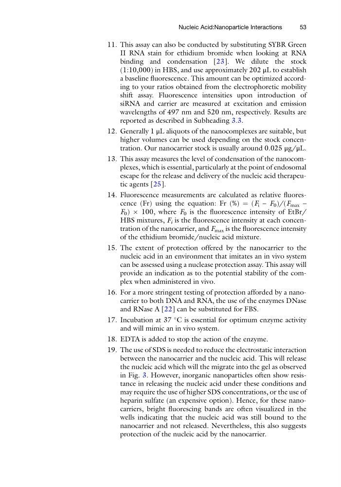
14. Fluorescence measurements are calculated as relative fluorescence (Fr) using the equation: Fr (%) ¼ (F i – F 0 )/(F max –
F 0 ) Â 100, where F 0 is the fluorescence intensity of EtBr/
HBS mixtures, F i is the fluorescence intensity at each concentration of the nanocarrier, and F max is the fluorescence intensity
of the ethidium bromide/nucleic acid mixture.
15. The extent of protection offered by the nanocarrier to the
nucleic acid in an environment that imitates an in vivo system
can be assessed using a nuclease protection assay. This assay will
provide an indication as to the potential stability of the complex when administered in vivo.
16. For a more stringent testing of protection afforded by a nanocarrier to both DNA and RNA, the use of the enzymes DNase
and RNase A [22] can be substituted for FBS.
17. Incubation at 37
C is essential for optimum enzyme activity
and will mimic an in vivo system.
18. EDTA is added to stop the action of the enzyme.
19. The use of SDS is needed to reduce the electrostatic interaction
between the nanocarrier and the nucleic acid. This will release
the nucleic acid which will the migrate into the gel as observed
in Fig. 3. However, inorganic nanoparticles often show resistance in releasing the nucleic acid under these conditions and
may require the use of higher SDS concentrations, or the use of
heparin sulfate (an expensive option). Hence, for these nanocarriers, bright fluorescing bands are often visualized in the
wells indicating that the nucleic acid was still bound to the
nanocarrier and not released. Nevertheless, this also suggests
protection of the nucleic acid by the nanocarrier.
Nucleic Acid:Nanoparticle Interactions
53
II RNA stain for ethidium bromide when looking at RNA
binding and condensation [23]. We dilute the stock
(1:10,000) in HBS, and use approximately 202 μL to establish
a baseline fluorescence. This amount can be optimized according to your ratios obtained from the electrophoretic mobility
shift assay. Fluorescence intensities upon introduction of
siRNA and carrier are measured at excitation and emission
wavelengths of 497 nm and 520 nm, respectively. Results are
reported as described in Subheading 3.3.
12. Generally 1 μL aliquots of the nanocomplexes are suitable, but
higher volumes can be used depending on the stock concentration. Our nanocarrier stock is usually around 0.025 μg/μL.
13. This assay measures the level of condensation of the nanocomplexes, which is essential, particularly at the point of endosomal
escape for the release and delivery of the nucleic acid therapeutic agents [25].
14. Fluorescence measurements are calculated as relative fluorescence (Fr) using the equation: Fr (%) ¼ (F i – F 0 )/(F max –
F 0 ) Â 100, where F 0 is the fluorescence intensity of EtBr/
HBS mixtures, F i is the fluorescence intensity at each concentration of the nanocarrier, and F max is the fluorescence intensity
of the ethidium bromide/nucleic acid mixture.
15. The extent of protection offered by the nanocarrier to the
nucleic acid in an environment that imitates an in vivo system
can be assessed using a nuclease protection assay. This assay will
provide an indication as to the potential stability of the complex when administered in vivo.
16. For a more stringent testing of protection afforded by a nanocarrier to both DNA and RNA, the use of the enzymes DNase
and RNase A [22] can be substituted for FBS.
17. Incubation at 37
C is essential for optimum enzyme activity
and will mimic an in vivo system.
18. EDTA is added to stop the action of the enzyme.
19. The use of SDS is needed to reduce the electrostatic interaction
between the nanocarrier and the nucleic acid. This will release
the nucleic acid which will the migrate into the gel as observed
in Fig. 3. However, inorganic nanoparticles often show resistance in releasing the nucleic acid under these conditions and
may require the use of higher SDS concentrations, or the use of
heparin sulfate (an expensive option). Hence, for these nanocarriers, bright fluorescing bands are often visualized in the
wells indicating that the nucleic acid was still bound to the
nanocarrier and not released. Nevertheless, this also suggests
protection of the nucleic acid by the nanocarrier.
Nucleic Acid:Nanoparticle Interactions
53