

4. Chill the cells on ice and proceed with FACS immediately.
5. Use the non-transfected K562 or Raji cells as a gating control
for FACS.
6. Sort the GFP
+ K562 or Raji cells into a 5-mL round-bottom
tube with a cap containing 500 μL of culture medium.
7. Centrifuge at 500 Â g for 5 min at room temperature.
8. Discard the supernatant, resuspend the pellet with the culture
medium, and transfer the sorted cells into a culture vessel (see
Note 12).
9. Incubate the cells at 37
C, 5% CO 2 .
10. On day 3 post-FAC sorting, transfer the cells to a 25-cm
2 cell
culture flask.
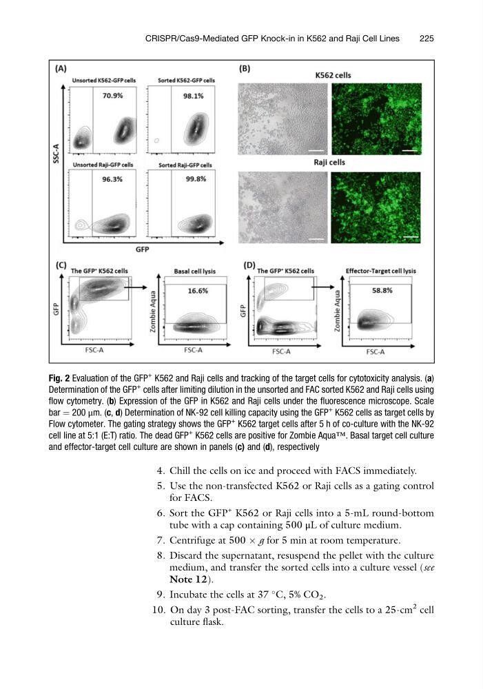
Fig. 2 Evaluation of the GFP
+ K562 and Raji cells and tracking of the target cells for cytotoxicity analysis. (a)
Determination of the GFP
+
cells after limiting dilution in the unsorted and FAC sorted K562 and Raji cells using
flow cytometry. (b) Expression of the GFP in K562 and Raji cells under the fluorescence microscope. Scale
bar ¼ 200 μm. (c, d) Determination of NK-92 cell killing capacity using the GFP
+
K562 cells as target cells by
Flow cytometer. The gating strategy shows the GFP
+
K562 target cells after 5 h of co-culture with the NK-92
cell line at 5:1 (E:T) ratio. The dead GFP
+
K562 cells are positive for Zombie Aqua™. Basal target cell culture
and effector-target cell culture are shown in panels (c) and (d), respectively
CRISPR/Cas9-Mediated GFP Knock-in in K562 and Raji Cell Lines
225
5. Use the non-transfected K562 or Raji cells as a gating control
for FACS.
6. Sort the GFP
+ K562 or Raji cells into a 5-mL round-bottom
tube with a cap containing 500 μL of culture medium.
7. Centrifuge at 500 Â g for 5 min at room temperature.
8. Discard the supernatant, resuspend the pellet with the culture
medium, and transfer the sorted cells into a culture vessel (see
Note 12).
9. Incubate the cells at 37
C, 5% CO 2 .
10. On day 3 post-FAC sorting, transfer the cells to a 25-cm
2 cell
culture flask.
Fig. 2 Evaluation of the GFP
+ K562 and Raji cells and tracking of the target cells for cytotoxicity analysis. (a)
Determination of the GFP
+
cells after limiting dilution in the unsorted and FAC sorted K562 and Raji cells using
flow cytometry. (b) Expression of the GFP in K562 and Raji cells under the fluorescence microscope. Scale
bar ¼ 200 μm. (c, d) Determination of NK-92 cell killing capacity using the GFP
+
K562 cells as target cells by
Flow cytometer. The gating strategy shows the GFP
+
K562 target cells after 5 h of co-culture with the NK-92
cell line at 5:1 (E:T) ratio. The dead GFP
+
K562 cells are positive for Zombie Aqua™. Basal target cell culture
and effector-target cell culture are shown in panels (c) and (d), respectively
CRISPR/Cas9-Mediated GFP Knock-in in K562 and Raji Cell Lines
225