

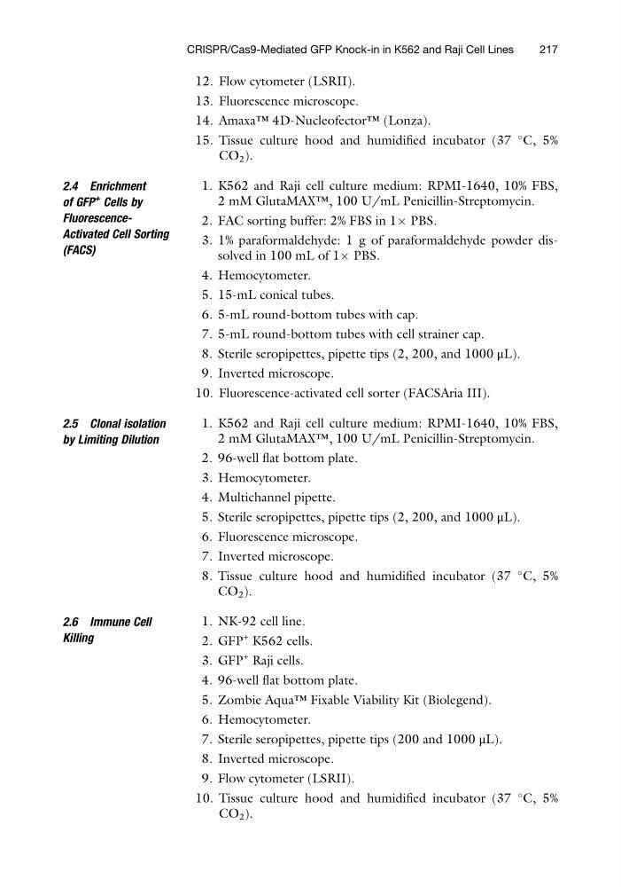
12. Flow cytometer (LSRII).
13. Fluorescence microscope.
14. Amaxa™ 4D-Nucleofector™ (Lonza).
15. Tissue culture hood and humidified incubator (37
C, 5%
CO 2 ).
2.4 Enrichment
of GFP
+ Cells by
FluorescenceActivated Cell Sorting
(FACS)
1. K562 and Raji cell culture medium: RPMI-1640, 10% FBS,
2 mM GlutaMAX™, 100 U/mL Penicillin-Streptomycin.
2. FAC sorting buffer: 2% FBS in 1Â PBS.
3. 1% paraformaldehyde: 1 g of paraformaldehyde powder dissolved in 100 mL of 1Â PBS.
4. Hemocytometer.
5. 15-mL conical tubes.
6. 5-mL round-bottom tubes with cap.
7. 5-mL round-bottom tubes with cell strainer cap.
8. Sterile seropipettes, pipette tips (2, 200, and 1000 μL).
9. Inverted microscope.
10. Fluorescence-activated cell sorter (FACSAria III).
2.5 Clonal isolation
by Limiting Dilution
1. K562 and Raji cell culture medium: RPMI-1640, 10% FBS,
2 mM GlutaMAX™, 100 U/mL Penicillin-Streptomycin.
2. 96-well flat bottom plate.
3. Hemocytometer.
4. Multichannel pipette.
5. Sterile seropipettes, pipette tips (2, 200, and 1000 μL).
6. Fluorescence microscope.
7. Inverted microscope.
8. Tissue culture hood and humidified incubator (37
C, 5%
CO 2 ).
2.6 Immune Cell
Killing
1. NK-92 cell line.
2. GFP
+ K562 cells.
3. GFP
+ Raji cells.
4. 96-well flat bottom plate.
5. Zombie Aqua™ Fixable Viability Kit (Biolegend).
6. Hemocytometer.
7. Sterile seropipettes, pipette tips (200 and 1000 μL).
8. Inverted microscope.
9. Flow cytometer (LSRII).
10. Tissue culture hood and humidified incubator (37
C, 5%
CO 2 ).
CRISPR/Cas9-Mediated GFP Knock-in in K562 and Raji Cell Lines
217
13. Fluorescence microscope.
14. Amaxa™ 4D-Nucleofector™ (Lonza).
15. Tissue culture hood and humidified incubator (37
C, 5%
CO 2 ).
2.4 Enrichment
of GFP
+ Cells by
FluorescenceActivated Cell Sorting
(FACS)
1. K562 and Raji cell culture medium: RPMI-1640, 10% FBS,
2 mM GlutaMAX™, 100 U/mL Penicillin-Streptomycin.
2. FAC sorting buffer: 2% FBS in 1Â PBS.
3. 1% paraformaldehyde: 1 g of paraformaldehyde powder dissolved in 100 mL of 1Â PBS.
4. Hemocytometer.
5. 15-mL conical tubes.
6. 5-mL round-bottom tubes with cap.
7. 5-mL round-bottom tubes with cell strainer cap.
8. Sterile seropipettes, pipette tips (2, 200, and 1000 μL).
9. Inverted microscope.
10. Fluorescence-activated cell sorter (FACSAria III).
2.5 Clonal isolation
by Limiting Dilution
1. K562 and Raji cell culture medium: RPMI-1640, 10% FBS,
2 mM GlutaMAX™, 100 U/mL Penicillin-Streptomycin.
2. 96-well flat bottom plate.
3. Hemocytometer.
4. Multichannel pipette.
5. Sterile seropipettes, pipette tips (2, 200, and 1000 μL).
6. Fluorescence microscope.
7. Inverted microscope.
8. Tissue culture hood and humidified incubator (37
C, 5%
CO 2 ).
2.6 Immune Cell
Killing
1. NK-92 cell line.
2. GFP
+ K562 cells.
3. GFP
+ Raji cells.
4. 96-well flat bottom plate.
5. Zombie Aqua™ Fixable Viability Kit (Biolegend).
6. Hemocytometer.
7. Sterile seropipettes, pipette tips (200 and 1000 μL).
8. Inverted microscope.
9. Flow cytometer (LSRII).
10. Tissue culture hood and humidified incubator (37
C, 5%
CO 2 ).
CRISPR/Cas9-Mediated GFP Knock-in in K562 and Raji Cell Lines
217