

3.5 Hemolysis Assay
The following assay is only conducted on bacterial vectors that
contain listeriolysin O (LLO) and assesses the activity of recombinantly produced LLO via the ability to disrupt red blood cells
(Fig. 4).
1. Sonicate the pellets saved during the DNA/protein release test
described above after resuspending in 1 mL of PBS at an
amplitude of 20% for 30 s with Branson 450D sonifier.
2. Following sonication, centrifuge the samples at 16,000 rpm
using a microfuge for 10 min at 4
C.
3. Following centrifugation, mix 100 μL of supernatant with
900 μL of 5% red blood cells in PBS buffer containing 6 mM
cysteine at two separate pH values of 5.15 and 7.4. Include red
blood cells that have not been mixed with bacterial lysate as a
control for 0% hemolysis. Add sodium dodecyl sulfate (SDS) to
the red blood cells to a concentration of 0.1% to achieve 100%
hemolysis as a positive control.
4. Incubate the mixture at 37
C for an hour.
5. Quantify LLO activity by measuring absorbance at 541 nm as
compared with a standard (centrifuged 5% red blood cell in a
corresponding PBS buffer) to calculate % red blood cell (RBC)
lysis. The calculation to be used is as follows:
%Hemolysis ¼ Sample À negative control
ð
Þ = positive control À negative control
ð
Þ ∗100%
Inoculation (10 mL)
to 0.4 OD 600nm
(2.5% (v/v),37 °C, 250 rpm)
Centrifuge for 10 min
(13000 rpm, 4 °C)
IPTG injection
Standardize to 0.5 OD 600nm
(100 µM)
Transfer supernatant to
15 mL centrifuge tube
Measure DNA (260 nm) and Protein (280 nM)
Sonicate 1 mL of supernatant
(20% amplitude, 30 seconds)
Centrifuge for 10 min
(16000 rpm, 4 °C)
Incubate for 1 hour
(37 °C)
Synergy 4 multi-mode
microplate reader
Mix supernatant (100 µL) with RBC in
buffer (pH 5.15, 7.4)
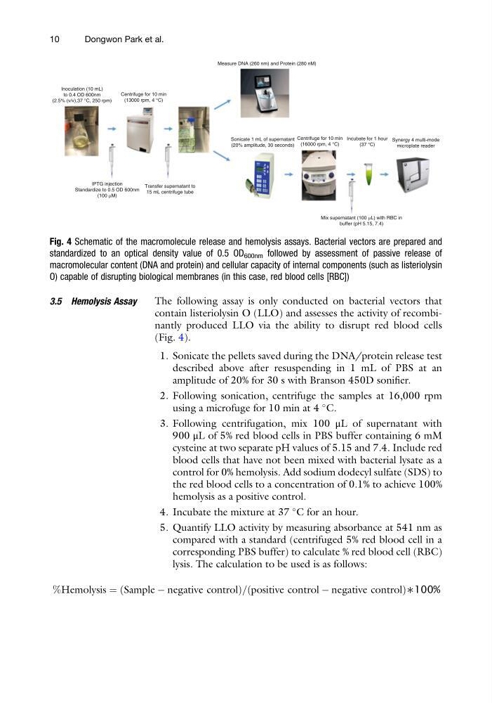
Fig. 4 Schematic of the macromolecule release and hemolysis assays. Bacterial vectors are prepared and
standardized to an optical density value of 0.5 OD 600nm followed by assessment of passive release of
macromolecular content (DNA and protein) and cellular capacity of internal components (such as listeriolysin
O) capable of disrupting biological membranes (in this case, red blood cells [RBC])
10
Dongwon Park et al.
The following assay is only conducted on bacterial vectors that
contain listeriolysin O (LLO) and assesses the activity of recombinantly produced LLO via the ability to disrupt red blood cells
(Fig. 4).
1. Sonicate the pellets saved during the DNA/protein release test
described above after resuspending in 1 mL of PBS at an
amplitude of 20% for 30 s with Branson 450D sonifier.
2. Following sonication, centrifuge the samples at 16,000 rpm
using a microfuge for 10 min at 4
C.
3. Following centrifugation, mix 100 μL of supernatant with
900 μL of 5% red blood cells in PBS buffer containing 6 mM
cysteine at two separate pH values of 5.15 and 7.4. Include red
blood cells that have not been mixed with bacterial lysate as a
control for 0% hemolysis. Add sodium dodecyl sulfate (SDS) to
the red blood cells to a concentration of 0.1% to achieve 100%
hemolysis as a positive control.
4. Incubate the mixture at 37
C for an hour.
5. Quantify LLO activity by measuring absorbance at 541 nm as
compared with a standard (centrifuged 5% red blood cell in a
corresponding PBS buffer) to calculate % red blood cell (RBC)
lysis. The calculation to be used is as follows:
%Hemolysis ¼ Sample À negative control
ð
Þ = positive control À negative control
ð
Þ ∗100%
Inoculation (10 mL)
to 0.4 OD 600nm
(2.5% (v/v),37 °C, 250 rpm)
Centrifuge for 10 min
(13000 rpm, 4 °C)
IPTG injection
Standardize to 0.5 OD 600nm
(100 µM)
Transfer supernatant to
15 mL centrifuge tube
Measure DNA (260 nm) and Protein (280 nM)
Sonicate 1 mL of supernatant
(20% amplitude, 30 seconds)
Centrifuge for 10 min
(16000 rpm, 4 °C)
Incubate for 1 hour
(37 °C)
Synergy 4 multi-mode
microplate reader
Mix supernatant (100 µL) with RBC in
buffer (pH 5.15, 7.4)
Fig. 4 Schematic of the macromolecule release and hemolysis assays. Bacterial vectors are prepared and
standardized to an optical density value of 0.5 OD 600nm followed by assessment of passive release of
macromolecular content (DNA and protein) and cellular capacity of internal components (such as listeriolysin
O) capable of disrupting biological membranes (in this case, red blood cells [RBC])
10
Dongwon Park et al.