

9 Mössbauer Spectroscopic Studies on Atomic Diffusion in Materials
487
(a)
(b)
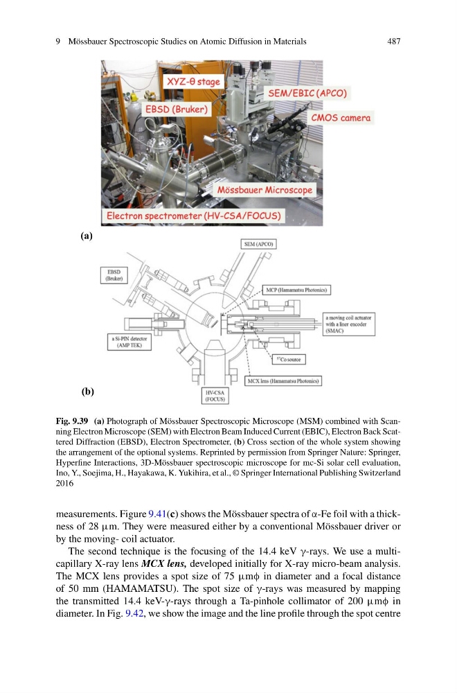
Fig. 9.39 (a) Photograph of Mössbauer Spectroscopic Microscope (MSM) combined with Scanning Electron Microscope (SEM) with Electron Beam Induced Current (EBIC), Electron Back Scattered Diffraction (EBSD), Electron Spectrometer, (b) Cross section of the whole system showing
the arrangement of the optional systems. Reprinted by permission from Springer Nature: Springer,
Hyperfine Interactions, 3D-Mössbauer spectroscopic microscope for mc-Si solar cell evaluation,
Ino, Y., Soejima, H., Hayakawa, K. Yukihira, et al., © Springer International Publishing Switzerland
2016
measurements. Figure 9.41(c) shows the Mössbauer spectra of α-Fe foil with a thickness of 28 μm. They were measured either by a conventional Mössbauer driver or
by the moving- coil actuator.
The second technique is the focusing of the 14.4 keV γ-rays. We use a multicapillary X-ray lens MCX lens, developed initially for X-ray micro-beam analysis.
The MCX lens provides a spot size of 75 μmφ in diameter and a focal distance
of 50 mm (HAMAMATSU). The spot size of γ-rays was measured by mapping
the transmitted 14.4 keV-γ-rays through a Ta-pinhole collimator of 200 μmφ in
diameter. In Fig. 9.42, we show the image and the line profile through the spot centre
487
(a)
(b)
Fig. 9.39 (a) Photograph of Mössbauer Spectroscopic Microscope (MSM) combined with Scanning Electron Microscope (SEM) with Electron Beam Induced Current (EBIC), Electron Back Scattered Diffraction (EBSD), Electron Spectrometer, (b) Cross section of the whole system showing
the arrangement of the optional systems. Reprinted by permission from Springer Nature: Springer,
Hyperfine Interactions, 3D-Mössbauer spectroscopic microscope for mc-Si solar cell evaluation,
Ino, Y., Soejima, H., Hayakawa, K. Yukihira, et al., © Springer International Publishing Switzerland
2016
measurements. Figure 9.41(c) shows the Mössbauer spectra of α-Fe foil with a thickness of 28 μm. They were measured either by a conventional Mössbauer driver or
by the moving- coil actuator.
The second technique is the focusing of the 14.4 keV γ-rays. We use a multicapillary X-ray lens MCX lens, developed initially for X-ray micro-beam analysis.
The MCX lens provides a spot size of 75 μmφ in diameter and a focal distance
of 50 mm (HAMAMATSU). The spot size of γ-rays was measured by mapping
the transmitted 14.4 keV-γ-rays through a Ta-pinhole collimator of 200 μmφ in
diameter. In Fig. 9.42, we show the image and the line profile through the spot centre