

9 Mössbauer Spectroscopic Studies on Atomic Diffusion in Materials
459
jump mechanism, we would like to observe the point defects, which jump adjacent
to the nuclear probes, and subsequently, exchange the lattice positions, and finally
lead to the atomic jumps of
57 Fe. However, the concentration of such point defects
will be rather low on the thermal equilibrium; for instance, only below 10
18 /cm
3 in
metals even just below the melting point. It may be possible, on the other hand, to
create an experimental condition where the concentration around
57 Fe atoms could
be orders of magnitude higher than that on the thermal equilibrium, i.e., under irradiations by highly energetic particles such as neutrons, electrons and heavy ions at
low temperature.
In this section, we explain “a thermal scanning method” in Mössbauer spectroscopy, which enables us to detect the recovery stages of the point defects in
pure Fe after low-temperature irradiations. Concerning the migration enthalpies of
vacancies, interstitials, and also carbon impurities in pure Fe, there were intensive
discussions among different research groups [36, 37] during the 1970s and 1980s.
Accordingly, we developed a thermal scanning method and applied for the pure Fe
samples carburized [38], or irradiated by energetic neutrons and electrons at low
temperatures [39], to clarify the kinetics of the recovery stages of the point defects
in pure iron.
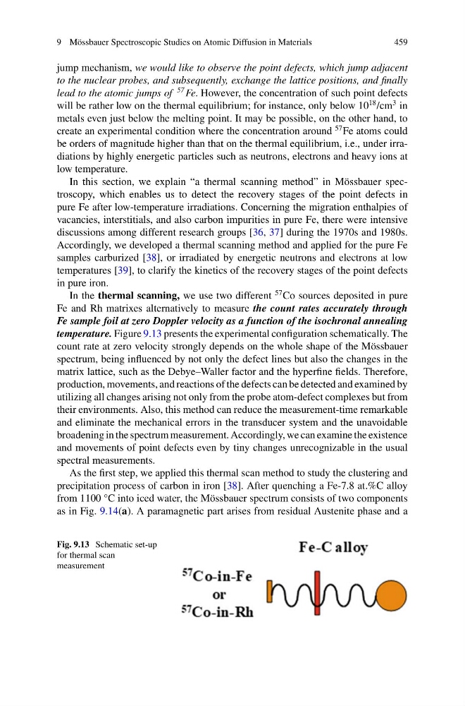
In the thermal scanning, we use two different
57 Co sources deposited in pure
Fe and Rh matrixes alternatively to measure the count rates accurately through
Fe sample foil at zero Doppler velocity as a function of the isochronal annealing
temperature. Figure 9.13 presents the experimental configuration schematically. The
count rate at zero velocity strongly depends on the whole shape of the Mössbauer
spectrum, being influenced by not only the defect lines but also the changes in the
matrix lattice, such as the Debye–Waller factor and the hyperfine fields. Therefore,
production, movements, and reactions of the defects can be detected and examined by
utilizing all changes arising not only from the probe atom-defect complexes but from
their environments. Also, this method can reduce the measurement-time remarkable
and eliminate the mechanical errors in the transducer system and the unavoidable
broadening in the spectrum measurement. Accordingly, we can examine the existence
and movements of point defects even by tiny changes unrecognizable in the usual
spectral measurements.
As the first step, we applied this thermal scan method to study the clustering and
precipitation process of carbon in iron [38]. After quenching a Fe-7.8 at.%C alloy
from 1100 °C into iced water, the Mössbauer spectrum consists of two components
as in Fig. 9.14(a). A paramagnetic part arises from residual Austenite phase and a
Fig. 9.13 Schematic set-up
for thermal scan
measurement
459
jump mechanism, we would like to observe the point defects, which jump adjacent
to the nuclear probes, and subsequently, exchange the lattice positions, and finally
lead to the atomic jumps of
57 Fe. However, the concentration of such point defects
will be rather low on the thermal equilibrium; for instance, only below 10
18 /cm
3 in
metals even just below the melting point. It may be possible, on the other hand, to
create an experimental condition where the concentration around
57 Fe atoms could
be orders of magnitude higher than that on the thermal equilibrium, i.e., under irradiations by highly energetic particles such as neutrons, electrons and heavy ions at
low temperature.
In this section, we explain “a thermal scanning method” in Mössbauer spectroscopy, which enables us to detect the recovery stages of the point defects in
pure Fe after low-temperature irradiations. Concerning the migration enthalpies of
vacancies, interstitials, and also carbon impurities in pure Fe, there were intensive
discussions among different research groups [36, 37] during the 1970s and 1980s.
Accordingly, we developed a thermal scanning method and applied for the pure Fe
samples carburized [38], or irradiated by energetic neutrons and electrons at low
temperatures [39], to clarify the kinetics of the recovery stages of the point defects
in pure iron.
In the thermal scanning, we use two different
57 Co sources deposited in pure
Fe and Rh matrixes alternatively to measure the count rates accurately through
Fe sample foil at zero Doppler velocity as a function of the isochronal annealing
temperature. Figure 9.13 presents the experimental configuration schematically. The
count rate at zero velocity strongly depends on the whole shape of the Mössbauer
spectrum, being influenced by not only the defect lines but also the changes in the
matrix lattice, such as the Debye–Waller factor and the hyperfine fields. Therefore,
production, movements, and reactions of the defects can be detected and examined by
utilizing all changes arising not only from the probe atom-defect complexes but from
their environments. Also, this method can reduce the measurement-time remarkable
and eliminate the mechanical errors in the transducer system and the unavoidable
broadening in the spectrum measurement. Accordingly, we can examine the existence
and movements of point defects even by tiny changes unrecognizable in the usual
spectral measurements.
As the first step, we applied this thermal scan method to study the clustering and
precipitation process of carbon in iron [38]. After quenching a Fe-7.8 at.%C alloy
from 1100 °C into iced water, the Mössbauer spectrum consists of two components
as in Fig. 9.14(a). A paramagnetic part arises from residual Austenite phase and a
Fig. 9.13 Schematic set-up
for thermal scan
measurement