

362
P.-E. Lippens
can be attributed to the progressive formation of superparamagnetic α-Fe nanoparticles with a constant average diameter of about 3 nm. This result is consistent with
the extrusion of Fe atoms from FeSn 2 during the conversion reaction and the formation of α-Fe nanoparticles. The small size of the formed α-Fe particles explains the
absence of XRD peaks. These results indicate that the first lithiation of FeSn 2 based
electrodes should be considered as a restructuring step ending with the formation
of α-Fe/Li 7 Sn 2 nanocomposite, which is the real starting electrode material for the
reversible lithiation-delithiation cycles.
Decreasing the particle size or increasing the surface area of electrochemically
active materials are expected to improve the performance by enhancing the electrolyte impregnation, Li diffusion and electronic percolation. Nanostructured FeSn 2
particles were obtained by ball milling of FeSn 2 microparticles as confirmed by XRD
showing broad Bragg peaks. The
57 Fe and
119 Sn Mössbauer spectra are formed by a
single peak and a doublet, respectively, indicating the absence of hyperfine magnetic
field as expected with the small size and poor crystallinity of the FeSn 2 ground particles [126]. However, the values of the isomer shift and quadrupole splitting are close
to those of the antiferromagnetic FeSn 2 crystalline phase, showing that Fe and Sn
local environments are similar in both nanostructured and crystalline FeSn 2 . During
the first lithiation, the voltage curve of nanostructured FeSn 2 decreases smoothly in
contrast to the plateau observed for FeSn 2 microparticles, while
57 Fe and
119 Sn Mössbauer spectra both reflect the same conversion reaction as given by Eq. (7.32) [68].
Thus, nanostructured and crystalline FeSn 2 electrode materials are both transformed
into an α-Fe/Li 7 Sn 2 nanocomposite during the first lithiation.
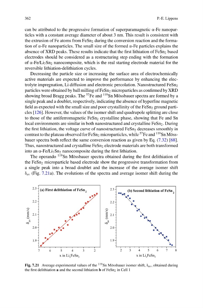
The operando
119 Sn Mössbauer spectra obtained during the first delithiation of
the FeSn 2 microparticle based electrode show the progressive transformation from
a single peak into a broad doublet and the increase of the average isomer shift
δ av (Fig. 7.21a). The evolutions of the spectra and average isomer shift during the
1.9
2.0
2.1
2.2
2.3
δ
δ
av (mm/s)
x in Li x FeSn 2
(a) First delithiation of FeSn 2
7
6
5
4
3
2
2
3
4
5
6
7
8
1.9
2.0
2.1
2.2
2.3
av (mm/s)
x in Li x FeSn 2
(b) Second lithiation of FeSn 2
Fig. 7.21 Average experimental values of the 119 Sn Mössbauer isomer shift, δ av , obtained during
the first delithiation a and the second lithiation b of FeSn 2 in Cell 1
P.-E. Lippens
can be attributed to the progressive formation of superparamagnetic α-Fe nanoparticles with a constant average diameter of about 3 nm. This result is consistent with
the extrusion of Fe atoms from FeSn 2 during the conversion reaction and the formation of α-Fe nanoparticles. The small size of the formed α-Fe particles explains the
absence of XRD peaks. These results indicate that the first lithiation of FeSn 2 based
electrodes should be considered as a restructuring step ending with the formation
of α-Fe/Li 7 Sn 2 nanocomposite, which is the real starting electrode material for the
reversible lithiation-delithiation cycles.
Decreasing the particle size or increasing the surface area of electrochemically
active materials are expected to improve the performance by enhancing the electrolyte impregnation, Li diffusion and electronic percolation. Nanostructured FeSn 2
particles were obtained by ball milling of FeSn 2 microparticles as confirmed by XRD
showing broad Bragg peaks. The
57 Fe and
119 Sn Mössbauer spectra are formed by a
single peak and a doublet, respectively, indicating the absence of hyperfine magnetic
field as expected with the small size and poor crystallinity of the FeSn 2 ground particles [126]. However, the values of the isomer shift and quadrupole splitting are close
to those of the antiferromagnetic FeSn 2 crystalline phase, showing that Fe and Sn
local environments are similar in both nanostructured and crystalline FeSn 2 . During
the first lithiation, the voltage curve of nanostructured FeSn 2 decreases smoothly in
contrast to the plateau observed for FeSn 2 microparticles, while
57 Fe and
119 Sn Mössbauer spectra both reflect the same conversion reaction as given by Eq. (7.32) [68].
Thus, nanostructured and crystalline FeSn 2 electrode materials are both transformed
into an α-Fe/Li 7 Sn 2 nanocomposite during the first lithiation.
The operando
119 Sn Mössbauer spectra obtained during the first delithiation of
the FeSn 2 microparticle based electrode show the progressive transformation from
a single peak into a broad doublet and the increase of the average isomer shift
δ av (Fig. 7.21a). The evolutions of the spectra and average isomer shift during the
1.9
2.0
2.1
2.2
2.3
δ
δ
av (mm/s)
x in Li x FeSn 2
(a) First delithiation of FeSn 2
7
6
5
4
3
2
2
3
4
5
6
7
8
1.9
2.0
2.1
2.2
2.3
av (mm/s)
x in Li x FeSn 2
(b) Second lithiation of FeSn 2
Fig. 7.21 Average experimental values of the 119 Sn Mössbauer isomer shift, δ av , obtained during
the first delithiation a and the second lithiation b of FeSn 2 in Cell 1