

4 From Small Molecules to Complex Systems: A Survey of Chemical …
211
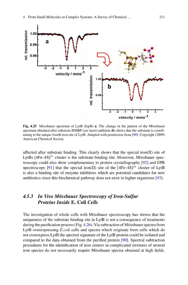
Fig. 4.25 Mössbauer spectrum of LytB (IspH) a. The change in the pattern of the Mössbauer
spectrum obtained after substrate HMBP (see inset) addition (b) shows that the substrate is coordinating to the unique fourth iron site of LytB. Adapted with permission from [90]. Copyright (2009)
American Chemical Society
affected after substrate binding. This clearly shows that the special iron(II) site of
LytBs [4Fe–4S]
2+ cluster is the substrate binding site. Moreover, Mössbauer spectroscopy could also show complementary to protein crystallography [92] and EPR
spectroscopy [91] that the special iron(II) site of the [4Fe–4S]
2+ cluster of LytB
is also a binding site of enzyme inhibitors which are potential candidates for new
antibiotics since this biochemical pathway does not exist in higher organisms [93].
4.5.3 In Vivo Mössbauer Spectroscopy of Iron-Sulfur
Proteins Inside E. Coli Cells
The investigation of whole cells with Mössbauer spectroscopy has shown that the
uniqueness of the substrate binding site in LytB is not a consequence of treatments
during the purification process (Fig. 4.26). Via subtraction of Mössbauer spectra from
LytB overexpressing E.coli cells and spectra which originate from cells which do
not overexpress LytB the spectral signature of the LytB protein could be isolated and
compared to the data obtained from the purified protein [90]. Spectral subtraction
procedures for the identification of iron centers in complicated mixtures of several
iron species do not necessarily require Mössbauer spectra obtained at high fields.
211
Fig. 4.25 Mössbauer spectrum of LytB (IspH) a. The change in the pattern of the Mössbauer
spectrum obtained after substrate HMBP (see inset) addition (b) shows that the substrate is coordinating to the unique fourth iron site of LytB. Adapted with permission from [90]. Copyright (2009)
American Chemical Society
affected after substrate binding. This clearly shows that the special iron(II) site of
LytBs [4Fe–4S]
2+ cluster is the substrate binding site. Moreover, Mössbauer spectroscopy could also show complementary to protein crystallography [92] and EPR
spectroscopy [91] that the special iron(II) site of the [4Fe–4S]
2+ cluster of LytB
is also a binding site of enzyme inhibitors which are potential candidates for new
antibiotics since this biochemical pathway does not exist in higher organisms [93].
4.5.3 In Vivo Mössbauer Spectroscopy of Iron-Sulfur
Proteins Inside E. Coli Cells
The investigation of whole cells with Mössbauer spectroscopy has shown that the
uniqueness of the substrate binding site in LytB is not a consequence of treatments
during the purification process (Fig. 4.26). Via subtraction of Mössbauer spectra from
LytB overexpressing E.coli cells and spectra which originate from cells which do
not overexpress LytB the spectral signature of the LytB protein could be isolated and
compared to the data obtained from the purified protein [90]. Spectral subtraction
procedures for the identification of iron centers in complicated mixtures of several
iron species do not necessarily require Mössbauer spectra obtained at high fields.