

9 Mössbauer Spectroscopic Studies on Atomic Diffusion in Materials
489
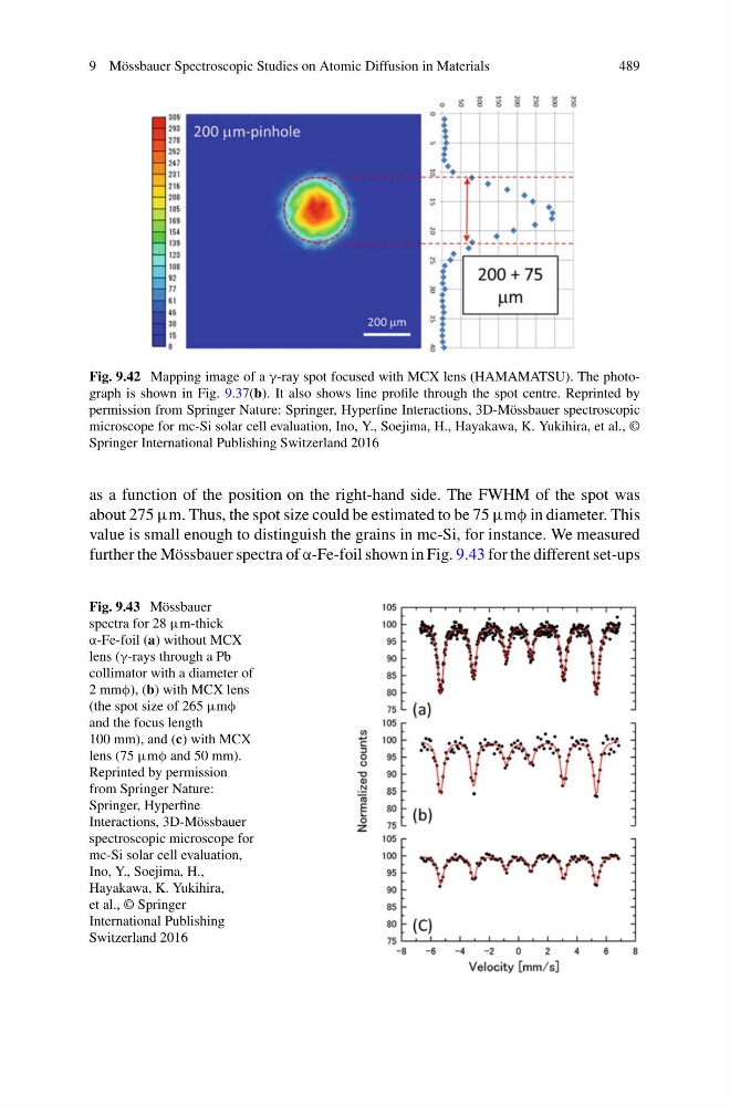
Fig. 9.42 Mapping image of a γ-ray spot focused with MCX lens (HAMAMATSU). The photograph is shown in Fig. 9.37(b). It also shows line profile through the spot centre. Reprinted by
permission from Springer Nature: Springer, Hyperfine Interactions, 3D-Mössbauer spectroscopic
microscope for mc-Si solar cell evaluation, Ino, Y., Soejima, H., Hayakawa, K. Yukihira, et al., ©
Springer International Publishing Switzerland 2016
as a function of the position on the right-hand side. The FWHM of the spot was
about 275 μm. Thus, the spot size could be estimated to be 75 μmφ in diameter. This
value is small enough to distinguish the grains in mc-Si, for instance. We measured
further the Mössbauer spectra of α-Fe-foil shown in Fig. 9.43 for the different set-ups
Fig. 9.43 Mössbauer
spectra for 28 μm-thick
α-Fe-foil (a) without MCX
lens (γ-rays through a Pb
collimator with a diameter of
2 mmφ), (b) with MCX lens
(the spot size of 265 μmφ
and the focus length
100 mm), and (c) with MCX
lens (75 μmφ and 50 mm).
Reprinted by permission
from Springer Nature:
Springer, Hyperfine
Interactions, 3D-Mössbauer
spectroscopic microscope for
mc-Si solar cell evaluation,
Ino, Y., Soejima, H.,
Hayakawa, K. Yukihira,
et al., © Springer
International Publishing
Switzerland 2016
489
Fig. 9.42 Mapping image of a γ-ray spot focused with MCX lens (HAMAMATSU). The photograph is shown in Fig. 9.37(b). It also shows line profile through the spot centre. Reprinted by
permission from Springer Nature: Springer, Hyperfine Interactions, 3D-Mössbauer spectroscopic
microscope for mc-Si solar cell evaluation, Ino, Y., Soejima, H., Hayakawa, K. Yukihira, et al., ©
Springer International Publishing Switzerland 2016
as a function of the position on the right-hand side. The FWHM of the spot was
about 275 μm. Thus, the spot size could be estimated to be 75 μmφ in diameter. This
value is small enough to distinguish the grains in mc-Si, for instance. We measured
further the Mössbauer spectra of α-Fe-foil shown in Fig. 9.43 for the different set-ups
Fig. 9.43 Mössbauer
spectra for 28 μm-thick
α-Fe-foil (a) without MCX
lens (γ-rays through a Pb
collimator with a diameter of
2 mmφ), (b) with MCX lens
(the spot size of 265 μmφ
and the focus length
100 mm), and (c) with MCX
lens (75 μmφ and 50 mm).
Reprinted by permission
from Springer Nature:
Springer, Hyperfine
Interactions, 3D-Mössbauer
spectroscopic microscope for
mc-Si solar cell evaluation,
Ino, Y., Soejima, H.,
Hayakawa, K. Yukihira,
et al., © Springer
International Publishing
Switzerland 2016