

472
Y. Yoshida
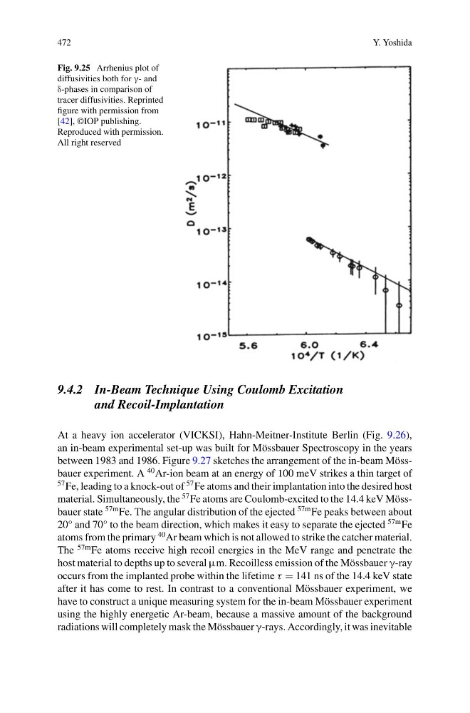
Fig. 9.25 Arrhenius plot of
diffusivities both for γ- and
δ-phases in comparison of
tracer diffusivities. Reprinted
figure with permission from
[42], ©IOP publishing.
Reproduced with permission.
All right reserved
9.4.2 In-Beam Technique Using Coulomb Excitation
and Recoil-Implantation
At a heavy ion accelerator (VICKSI), Hahn-Meitner-Institute Berlin (Fig. 9.26),
an in-beam experimental set-up was built for Mössbauer Spectroscopy in the years
between 1983 and 1986. Figure 9.27 sketches the arrangement of the in-beam Mössbauer experiment. A
40 Ar-ion beam at an energy of 100 meV strikes a thin target of
57 Fe, leading to a knock-out of
57 Fe atoms and their implantation into the desired host
material. Simultaneously, the
57 Fe atoms are Coulomb-excited to the 14.4 keV Mössbauer state
57m Fe. The angular distribution of the ejected
57m Fe peaks between about
20° and 70° to the beam direction, which makes it easy to separate the ejected
57m Fe
atoms from the primary
40 Ar beam which is not allowed to strike the catcher material.
The
57m Fe atoms receive high recoil energies in the MeV range and penetrate the
host material to depths up to several μm. Recoilless emission of the Mössbauer γ-ray
occurs from the implanted probe within the lifetime τ = 141 ns of the 14.4 keV state
after it has come to rest. In contrast to a conventional Mössbauer experiment, we
have to construct a unique measuring system for the in-beam Mössbauer experiment
using the highly energetic Ar-beam, because a massive amount of the background
radiations will completely mask the Mössbauer γ-rays. Accordingly, it was inevitable
Y. Yoshida
Fig. 9.25 Arrhenius plot of
diffusivities both for γ- and
δ-phases in comparison of
tracer diffusivities. Reprinted
figure with permission from
[42], ©IOP publishing.
Reproduced with permission.
All right reserved
9.4.2 In-Beam Technique Using Coulomb Excitation
and Recoil-Implantation
At a heavy ion accelerator (VICKSI), Hahn-Meitner-Institute Berlin (Fig. 9.26),
an in-beam experimental set-up was built for Mössbauer Spectroscopy in the years
between 1983 and 1986. Figure 9.27 sketches the arrangement of the in-beam Mössbauer experiment. A
40 Ar-ion beam at an energy of 100 meV strikes a thin target of
57 Fe, leading to a knock-out of
57 Fe atoms and their implantation into the desired host
material. Simultaneously, the
57 Fe atoms are Coulomb-excited to the 14.4 keV Mössbauer state
57m Fe. The angular distribution of the ejected
57m Fe peaks between about
20° and 70° to the beam direction, which makes it easy to separate the ejected
57m Fe
atoms from the primary
40 Ar beam which is not allowed to strike the catcher material.
The
57m Fe atoms receive high recoil energies in the MeV range and penetrate the
host material to depths up to several μm. Recoilless emission of the Mössbauer γ-ray
occurs from the implanted probe within the lifetime τ = 141 ns of the 14.4 keV state
after it has come to rest. In contrast to a conventional Mössbauer experiment, we
have to construct a unique measuring system for the in-beam Mössbauer experiment
using the highly energetic Ar-beam, because a massive amount of the background
radiations will completely mask the Mössbauer γ-rays. Accordingly, it was inevitable