

20
R. Rüffer and A. I. Chumakov
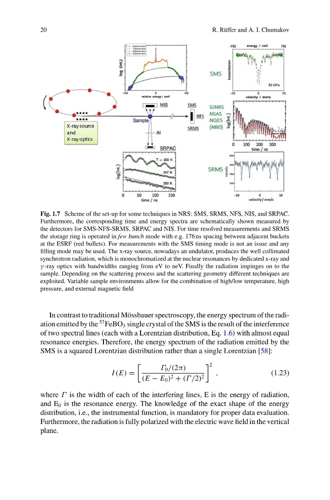
Fig. 1.7 Scheme of the set-up for some techniques in NRS: SMS, SRMS, NFS, NIS, and SRPAC.
Furthermore, the corresponding time and energy spectra are schematically shown measured by
the detectors for SMS-NFS-SRMS, SRPAC and NIS. For time resolved measurements and SRMS
the storage ring is operated in few bunch mode with e.g. 176 ns spacing between adjacent buckets
at the ESRF (red bullets). For measurements with the SMS timing mode is not an issue and any
filling mode may be used. The x-ray source, nowadays an undulator, produces the well collimated
synchrotron radiation, which is monochromatized at the nuclear resonances by dedicated x-ray and
γ -ray optics with bandwidths ranging from eV to neV. Finally the radiation impinges on to the
sample. Depending on the scattering process and the scattering geometry different techniques are
exploited. Variable sample environments allow for the combination of high/low temperature, high
pressure, and external magnetic field
In contrast to traditional Mössbauer spectroscopy, the energy spectrum of the radiation emitted by the
57 FeBO 3 single crystal of the SMS is the result of the interference
of two spectral lines (each with a Lorentzian distribution, Eq. 1.6) with almost equal
resonance energies. Therefore, the energy spectrum of the radiation emitted by the
SMS is a squared Lorentzian distribution rather than a single Lorentzian [58]:
I (E) =
Γ 0 /(2π)
(E − E 0 ) 2 + (Γ /2) 2
2
,
(1.23)
where Γ is the width of each of the interfering lines, E is the energy of radiation,
and E 0 is the resonance energy. The knowledge of the exact shape of the energy
distribution, i.e., the instrumental function, is mandatory for proper data evaluation.
Furthermore, the radiation is fully polarized with the electric wave field in the vertical
plane.
R. Rüffer and A. I. Chumakov
Fig. 1.7 Scheme of the set-up for some techniques in NRS: SMS, SRMS, NFS, NIS, and SRPAC.
Furthermore, the corresponding time and energy spectra are schematically shown measured by
the detectors for SMS-NFS-SRMS, SRPAC and NIS. For time resolved measurements and SRMS
the storage ring is operated in few bunch mode with e.g. 176 ns spacing between adjacent buckets
at the ESRF (red bullets). For measurements with the SMS timing mode is not an issue and any
filling mode may be used. The x-ray source, nowadays an undulator, produces the well collimated
synchrotron radiation, which is monochromatized at the nuclear resonances by dedicated x-ray and
γ -ray optics with bandwidths ranging from eV to neV. Finally the radiation impinges on to the
sample. Depending on the scattering process and the scattering geometry different techniques are
exploited. Variable sample environments allow for the combination of high/low temperature, high
pressure, and external magnetic field
In contrast to traditional Mössbauer spectroscopy, the energy spectrum of the radiation emitted by the
57 FeBO 3 single crystal of the SMS is the result of the interference
of two spectral lines (each with a Lorentzian distribution, Eq. 1.6) with almost equal
resonance energies. Therefore, the energy spectrum of the radiation emitted by the
SMS is a squared Lorentzian distribution rather than a single Lorentzian [58]:
I (E) =
Γ 0 /(2π)
(E − E 0 ) 2 + (Γ /2) 2
2
,
(1.23)
where Γ is the width of each of the interfering lines, E is the energy of radiation,
and E 0 is the resonance energy. The knowledge of the exact shape of the energy
distribution, i.e., the instrumental function, is mandatory for proper data evaluation.
Furthermore, the radiation is fully polarized with the electric wave field in the vertical
plane.