

212
V. Schünemann
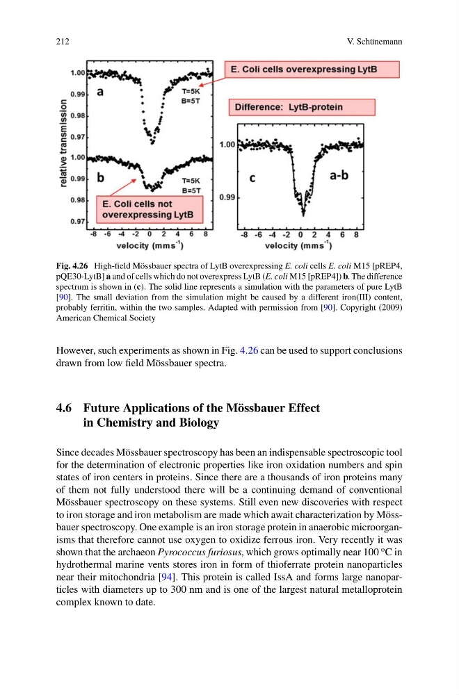
Fig. 4.26 High-field Mössbauer spectra of LytB overexpressing E. coli cells E. coli M15 [pREP4,
pQE30-LytB] a and of cells which do not overexpress LytB (E. coli M15 [pREP4]) b. The difference
spectrum is shown in (c). The solid line represents a simulation with the parameters of pure LytB
[90]. The small deviation from the simulation might be caused by a different iron(III) content,
probably ferritin, within the two samples. Adapted with permission from [90]. Copyright (2009)
American Chemical Society
However, such experiments as shown in Fig. 4.26 can be used to support conclusions
drawn from low field Mössbauer spectra.
4.6 Future Applications of the Mössbauer Effect
in Chemistry and Biology
Since decades Mössbauer spectroscopy has been an indispensable spectroscopic tool
for the determination of electronic properties like iron oxidation numbers and spin
states of iron centers in proteins. Since there are a thousands of iron proteins many
of them not fully understood there will be a continuing demand of conventional
Mössbauer spectroscopy on these systems. Still even new discoveries with respect
to iron storage and iron metabolism are made which await characterization by Mössbauer spectroscopy. One example is an iron storage protein in anaerobic microorganisms that therefore cannot use oxygen to oxidize ferrous iron. Very recently it was
shown that the archaeon Pyrococcus furiosus, which grows optimally near 100
o C in
hydrothermal marine vents stores iron in form of thioferrate protein nanoparticles
near their mitochondria [94]. This protein is called IssA and forms large nanoparticles with diameters up to 300 nm and is one of the largest natural metalloprotein
complex known to date.
V. Schünemann
Fig. 4.26 High-field Mössbauer spectra of LytB overexpressing E. coli cells E. coli M15 [pREP4,
pQE30-LytB] a and of cells which do not overexpress LytB (E. coli M15 [pREP4]) b. The difference
spectrum is shown in (c). The solid line represents a simulation with the parameters of pure LytB
[90]. The small deviation from the simulation might be caused by a different iron(III) content,
probably ferritin, within the two samples. Adapted with permission from [90]. Copyright (2009)
American Chemical Society
However, such experiments as shown in Fig. 4.26 can be used to support conclusions
drawn from low field Mössbauer spectra.
4.6 Future Applications of the Mössbauer Effect
in Chemistry and Biology
Since decades Mössbauer spectroscopy has been an indispensable spectroscopic tool
for the determination of electronic properties like iron oxidation numbers and spin
states of iron centers in proteins. Since there are a thousands of iron proteins many
of them not fully understood there will be a continuing demand of conventional
Mössbauer spectroscopy on these systems. Still even new discoveries with respect
to iron storage and iron metabolism are made which await characterization by Mössbauer spectroscopy. One example is an iron storage protein in anaerobic microorganisms that therefore cannot use oxygen to oxidize ferrous iron. Very recently it was
shown that the archaeon Pyrococcus furiosus, which grows optimally near 100
o C in
hydrothermal marine vents stores iron in form of thioferrate protein nanoparticles
near their mitochondria [94]. This protein is called IssA and forms large nanoparticles with diameters up to 300 nm and is one of the largest natural metalloprotein
complex known to date.