

208
V. Schünemann
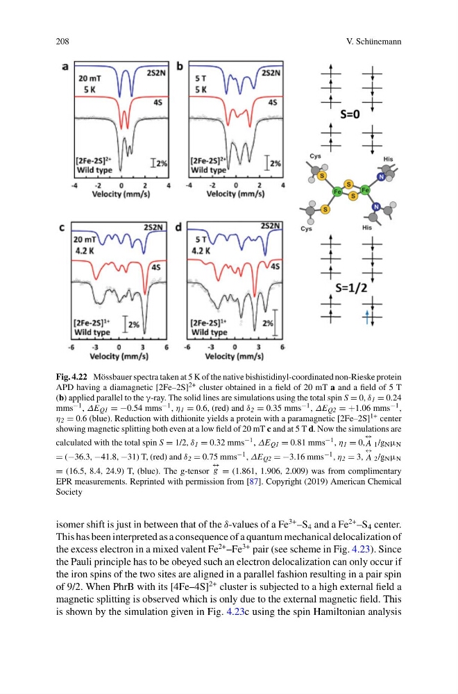
Fig. 4.22 Mössbauer spectra taken at 5 K of the native bishistidinyl-coordinated non-Rieske protein
APD having a diamagnetic [2Fe–2S] 2+ cluster obtained in a field of 20 mT a and a field of 5 T
(b) applied parallel to the γ-ray. The solid lines are simulations using the total spin S = 0, δ 1 = 0.24
mms −1 , ΔE Q1 = −0.54 mms −1 , η 1 = 0.6, (red) and δ 2 = 0.35 mms −1 , ΔE Q2 = +1.06 mms −1 ,
η 2 = 0.6 (blue). Reduction with dithionite yields a protein with a paramagnetic [2Fe–2S] 1+ center
showing magnetic splitting both even at a low field of 20 mT c and at 5 T d. Now the simulations are
calculated with the total spin S = 1/2, δ 1 = 0.32 mms −1 , ΔE Q1 = 0.81 mms −1 , η 1 = 0,
↔
A 1 /g N μ N
= (−36.3, −41.8, −31) T, (red) and δ 2 = 0.75 mms −1 , ΔE Q2 = −3.16 mms −1 , η 2 = 3,
↔
A 2 /g N μ N
= (16.5, 8.4, 24.9) T, (blue). The g-tensor
↔
g = (1.861, 1.906, 2.009) was from complimentary
EPR measurements. Reprinted with permission from [87]. Copyright (2019) American Chemical
Society
isomer shift is just in between that of the δ-values of a Fe
3+ –S 4 and a Fe
2+ –S 4 center.
This has been interpreted as a consequence of a quantum mechanical delocalization of
the excess electron in a mixed valent Fe
2+ –Fe
3+ pair (see scheme in Fig. 4.23). Since
the Pauli principle has to be obeyed such an electron delocalization can only occur if
the iron spins of the two sites are aligned in a parallel fashion resulting in a pair spin
of 9/2. When PhrB with its [4Fe–4S]
2+ cluster is subjected to a high external field a
magnetic splitting is observed which is only due to the external magnetic field. This
is shown by the simulation given in Fig. 4.23c using the spin Hamiltonian analysis
V. Schünemann
Fig. 4.22 Mössbauer spectra taken at 5 K of the native bishistidinyl-coordinated non-Rieske protein
APD having a diamagnetic [2Fe–2S] 2+ cluster obtained in a field of 20 mT a and a field of 5 T
(b) applied parallel to the γ-ray. The solid lines are simulations using the total spin S = 0, δ 1 = 0.24
mms −1 , ΔE Q1 = −0.54 mms −1 , η 1 = 0.6, (red) and δ 2 = 0.35 mms −1 , ΔE Q2 = +1.06 mms −1 ,
η 2 = 0.6 (blue). Reduction with dithionite yields a protein with a paramagnetic [2Fe–2S] 1+ center
showing magnetic splitting both even at a low field of 20 mT c and at 5 T d. Now the simulations are
calculated with the total spin S = 1/2, δ 1 = 0.32 mms −1 , ΔE Q1 = 0.81 mms −1 , η 1 = 0,
↔
A 1 /g N μ N
= (−36.3, −41.8, −31) T, (red) and δ 2 = 0.75 mms −1 , ΔE Q2 = −3.16 mms −1 , η 2 = 3,
↔
A 2 /g N μ N
= (16.5, 8.4, 24.9) T, (blue). The g-tensor
↔
g = (1.861, 1.906, 2.009) was from complimentary
EPR measurements. Reprinted with permission from [87]. Copyright (2019) American Chemical
Society
isomer shift is just in between that of the δ-values of a Fe
3+ –S 4 and a Fe
2+ –S 4 center.
This has been interpreted as a consequence of a quantum mechanical delocalization of
the excess electron in a mixed valent Fe
2+ –Fe
3+ pair (see scheme in Fig. 4.23). Since
the Pauli principle has to be obeyed such an electron delocalization can only occur if
the iron spins of the two sites are aligned in a parallel fashion resulting in a pair spin
of 9/2. When PhrB with its [4Fe–4S]
2+ cluster is subjected to a high external field a
magnetic splitting is observed which is only due to the external magnetic field. This
is shown by the simulation given in Fig. 4.23c using the spin Hamiltonian analysis