

204
V. Schünemann
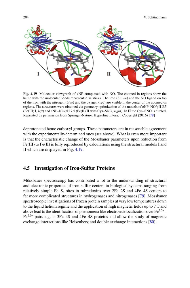
Fig. 4.19 Molecular viewgraph of cNP complexed with NO. The zoomed-in regions show the
heme with the molecular bonds represented as sticks. The iron (brown) and the NO ligand on top
of the iron with the nitrogen (blue) and the oxygen (red) are visible in the center of the zoomed-in
regions. The structures were obtained via geometry optimization of the models of cNP–NO/pH 5.5
(Fe(III) I, left) and cNP–NO/pH 7.5 (Fe(II) II with Cys–SNO, right). In II the Cys–SNO is circled.
Reprinted by permission from Springer-Nature: Hyperfine Interact. Copyright (2016) [78]
deprotonated heme carboxyl groups. These parameters are in reasonable agreement
with the experimentally-determined ones (see above). What is even more important
is that the characteristic change of the Mössbauer parameters upon reduction from
Fe(III) to Fe(II) is fully reproduced by calculations using the structural models I and
II which are displayed in Fig. 4.19.
4.5 Investigation of Iron-Sulfur Proteins
Mössbauer spectroscopy has contributed a lot to the understanding of structural
and electronic properties of iron-sulfur centers in biological systems ranging from
relatively simple Fe–S 4 sites in rubredoxins over 2Fe–2S and 4Fe–4S centers to
far more complicated structures in hydrogenases and nitrogenases [79]. Mössbauer
spectroscopic investigations of frozen protein samples at very low temperatures down
to the liquid helium regime and the application of high magnetic fields up to 7 T and
above lead to the identification of phenomena like electron delocalization over Fe
2.5+ –
Fe
2.5+ pairs e.g. in 3Fe–4S and 4Fe–4S proteins and allow the study of magnetic
exchange interactions like Heisenberg and double exchange interactions [80].
V. Schünemann
Fig. 4.19 Molecular viewgraph of cNP complexed with NO. The zoomed-in regions show the
heme with the molecular bonds represented as sticks. The iron (brown) and the NO ligand on top
of the iron with the nitrogen (blue) and the oxygen (red) are visible in the center of the zoomed-in
regions. The structures were obtained via geometry optimization of the models of cNP–NO/pH 5.5
(Fe(III) I, left) and cNP–NO/pH 7.5 (Fe(II) II with Cys–SNO, right). In II the Cys–SNO is circled.
Reprinted by permission from Springer-Nature: Hyperfine Interact. Copyright (2016) [78]
deprotonated heme carboxyl groups. These parameters are in reasonable agreement
with the experimentally-determined ones (see above). What is even more important
is that the characteristic change of the Mössbauer parameters upon reduction from
Fe(III) to Fe(II) is fully reproduced by calculations using the structural models I and
II which are displayed in Fig. 4.19.
4.5 Investigation of Iron-Sulfur Proteins
Mössbauer spectroscopy has contributed a lot to the understanding of structural
and electronic properties of iron-sulfur centers in biological systems ranging from
relatively simple Fe–S 4 sites in rubredoxins over 2Fe–2S and 4Fe–4S centers to
far more complicated structures in hydrogenases and nitrogenases [79]. Mössbauer
spectroscopic investigations of frozen protein samples at very low temperatures down
to the liquid helium regime and the application of high magnetic fields up to 7 T and
above lead to the identification of phenomena like electron delocalization over Fe
2.5+ –
Fe
2.5+ pairs e.g. in 3Fe–4S and 4Fe–4S proteins and allow the study of magnetic
exchange interactions like Heisenberg and double exchange interactions [80].