

4 From Small Molecules to Complex Systems: A Survey of Chemical …
175
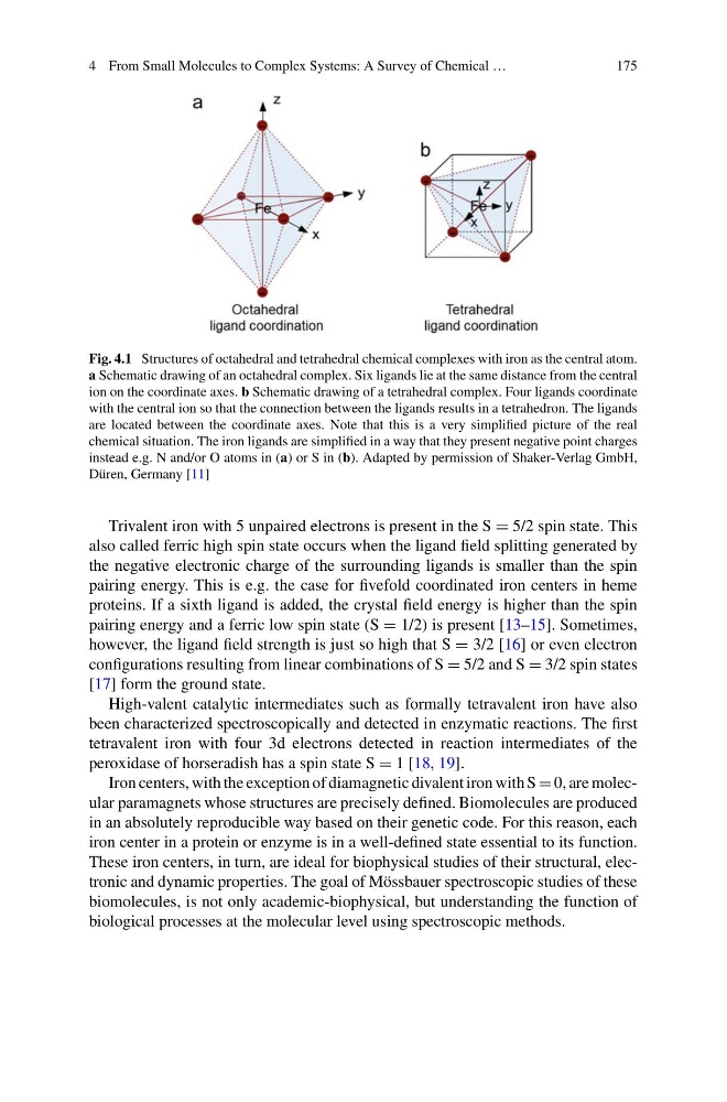
Fig. 4.1 Structures of octahedral and tetrahedral chemical complexes with iron as the central atom.
a Schematic drawing of an octahedral complex. Six ligands lie at the same distance from the central
ion on the coordinate axes. b Schematic drawing of a tetrahedral complex. Four ligands coordinate
with the central ion so that the connection between the ligands results in a tetrahedron. The ligands
are located between the coordinate axes. Note that this is a very simplified picture of the real
chemical situation. The iron ligands are simplified in a way that they present negative point charges
instead e.g. N and/or O atoms in (a) or S in (b). Adapted by permission of Shaker-Verlag GmbH,
Düren, Germany [11]
Trivalent iron with 5 unpaired electrons is present in the S = 5/2 spin state. This
also called ferric high spin state occurs when the ligand field splitting generated by
the negative electronic charge of the surrounding ligands is smaller than the spin
pairing energy. This is e.g. the case for fivefold coordinated iron centers in heme
proteins. If a sixth ligand is added, the crystal field energy is higher than the spin
pairing energy and a ferric low spin state (S = 1/2) is present [13–15]. Sometimes,
however, the ligand field strength is just so high that S = 3/2 [16] or even electron
configurations resulting from linear combinations of S = 5/2 and S = 3/2 spin states
[17] form the ground state.
High-valent catalytic intermediates such as formally tetravalent iron have also
been characterized spectroscopically and detected in enzymatic reactions. The first
tetravalent iron with four 3d electrons detected in reaction intermediates of the
peroxidase of horseradish has a spin state S = 1 [18, 19].
Iron centers, with the exception of diamagnetic divalent iron with S = 0, are molecular paramagnets whose structures are precisely defined. Biomolecules are produced
in an absolutely reproducible way based on their genetic code. For this reason, each
iron center in a protein or enzyme is in a well-defined state essential to its function.
These iron centers, in turn, are ideal for biophysical studies of their structural, electronic and dynamic properties. The goal of Mössbauer spectroscopic studies of these
biomolecules, is not only academic-biophysical, but understanding the function of
biological processes at the molecular level using spectroscopic methods.
175
Fig. 4.1 Structures of octahedral and tetrahedral chemical complexes with iron as the central atom.
a Schematic drawing of an octahedral complex. Six ligands lie at the same distance from the central
ion on the coordinate axes. b Schematic drawing of a tetrahedral complex. Four ligands coordinate
with the central ion so that the connection between the ligands results in a tetrahedron. The ligands
are located between the coordinate axes. Note that this is a very simplified picture of the real
chemical situation. The iron ligands are simplified in a way that they present negative point charges
instead e.g. N and/or O atoms in (a) or S in (b). Adapted by permission of Shaker-Verlag GmbH,
Düren, Germany [11]
Trivalent iron with 5 unpaired electrons is present in the S = 5/2 spin state. This
also called ferric high spin state occurs when the ligand field splitting generated by
the negative electronic charge of the surrounding ligands is smaller than the spin
pairing energy. This is e.g. the case for fivefold coordinated iron centers in heme
proteins. If a sixth ligand is added, the crystal field energy is higher than the spin
pairing energy and a ferric low spin state (S = 1/2) is present [13–15]. Sometimes,
however, the ligand field strength is just so high that S = 3/2 [16] or even electron
configurations resulting from linear combinations of S = 5/2 and S = 3/2 spin states
[17] form the ground state.
High-valent catalytic intermediates such as formally tetravalent iron have also
been characterized spectroscopically and detected in enzymatic reactions. The first
tetravalent iron with four 3d electrons detected in reaction intermediates of the
peroxidase of horseradish has a spin state S = 1 [18, 19].
Iron centers, with the exception of diamagnetic divalent iron with S = 0, are molecular paramagnets whose structures are precisely defined. Biomolecules are produced
in an absolutely reproducible way based on their genetic code. For this reason, each
iron center in a protein or enzyme is in a well-defined state essential to its function.
These iron centers, in turn, are ideal for biophysical studies of their structural, electronic and dynamic properties. The goal of Mössbauer spectroscopic studies of these
biomolecules, is not only academic-biophysical, but understanding the function of
biological processes at the molecular level using spectroscopic methods.