

3 Quantum Optical Phenomena in Nuclear Resonant Scattering
141
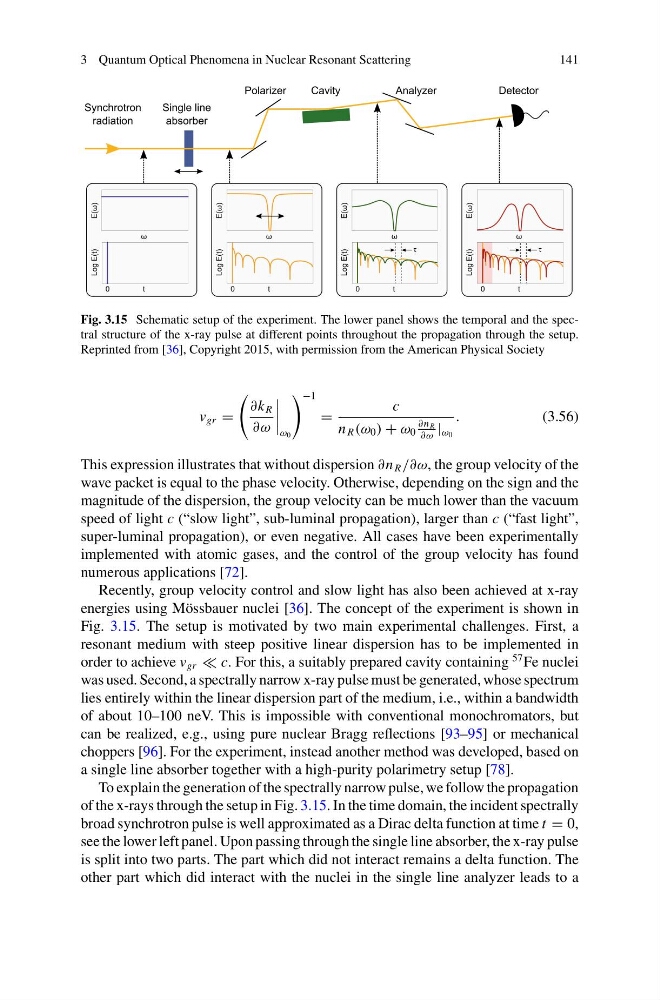
Fig. 3.15 Schematic setup of the experiment. The lower panel shows the temporal and the spectral structure of the x-ray pulse at different points throughout the propagation through the setup.
Reprinted from [36], Copyright 2015, with permission from the American Physical Society
v gr =
∂k R
∂ω
ω 0
−1
=
c
n R (ω 0 ) + ω 0
∂n R
∂ω
| ω 0
.
(3.56)
This expression illustrates that without dispersion ∂n R /∂ω, the group velocity of the
wave packet is equal to the phase velocity. Otherwise, depending on the sign and the
magnitude of the dispersion, the group velocity can be much lower than the vacuum
speed of light c (“slow light”, sub-luminal propagation), larger than c (“fast light”,
super-luminal propagation), or even negative. All cases have been experimentally
implemented with atomic gases, and the control of the group velocity has found
numerous applications [72].
Recently, group velocity control and slow light has also been achieved at x-ray
energies using Mössbauer nuclei [36]. The concept of the experiment is shown in
Fig. 3.15. The setup is motivated by two main experimental challenges. First, a
resonant medium with steep positive linear dispersion has to be implemented in
order to achieve v gr c. For this, a suitably prepared cavity containing
57 Fe nuclei
was used. Second, a spectrally narrow x-ray pulse must be generated, whose spectrum
lies entirely within the linear dispersion part of the medium, i.e., within a bandwidth
of about 10–100 neV. This is impossible with conventional monochromators, but
can be realized, e.g., using pure nuclear Bragg reflections [93–95] or mechanical
choppers [96]. For the experiment, instead another method was developed, based on
a single line absorber together with a high-purity polarimetry setup [78].
To explain the generation of the spectrally narrow pulse, we follow the propagation
of the x-rays through the setup in Fig. 3.15. In the time domain, the incident spectrally
broad synchrotron pulse is well approximated as a Dirac delta function at time t = 0,
see the lower left panel. Upon passing through the single line absorber, the x-ray pulse
is split into two parts. The part which did not interact remains a delta function. The
other part which did interact with the nuclei in the single line analyzer leads to a
141
Fig. 3.15 Schematic setup of the experiment. The lower panel shows the temporal and the spectral structure of the x-ray pulse at different points throughout the propagation through the setup.
Reprinted from [36], Copyright 2015, with permission from the American Physical Society
v gr =
∂k R
∂ω
ω 0
−1
=
c
n R (ω 0 ) + ω 0
∂n R
∂ω
| ω 0
.
(3.56)
This expression illustrates that without dispersion ∂n R /∂ω, the group velocity of the
wave packet is equal to the phase velocity. Otherwise, depending on the sign and the
magnitude of the dispersion, the group velocity can be much lower than the vacuum
speed of light c (“slow light”, sub-luminal propagation), larger than c (“fast light”,
super-luminal propagation), or even negative. All cases have been experimentally
implemented with atomic gases, and the control of the group velocity has found
numerous applications [72].
Recently, group velocity control and slow light has also been achieved at x-ray
energies using Mössbauer nuclei [36]. The concept of the experiment is shown in
Fig. 3.15. The setup is motivated by two main experimental challenges. First, a
resonant medium with steep positive linear dispersion has to be implemented in
order to achieve v gr c. For this, a suitably prepared cavity containing
57 Fe nuclei
was used. Second, a spectrally narrow x-ray pulse must be generated, whose spectrum
lies entirely within the linear dispersion part of the medium, i.e., within a bandwidth
of about 10–100 neV. This is impossible with conventional monochromators, but
can be realized, e.g., using pure nuclear Bragg reflections [93–95] or mechanical
choppers [96]. For the experiment, instead another method was developed, based on
a single line absorber together with a high-purity polarimetry setup [78].
To explain the generation of the spectrally narrow pulse, we follow the propagation
of the x-rays through the setup in Fig. 3.15. In the time domain, the incident spectrally
broad synchrotron pulse is well approximated as a Dirac delta function at time t = 0,
see the lower left panel. Upon passing through the single line absorber, the x-ray pulse
is split into two parts. The part which did not interact remains a delta function. The
other part which did interact with the nuclei in the single line analyzer leads to a