

used in these experiments resulted in hydrogels with flow properties resembling
those of the peptide hydrogels. Therefore, differences in cell behavior in different
matrices could not be due to differences in the mechanical properties of the
hydrogels.
4 In Vivo Studies
Despite the promising results reported so far in in vitro studies, it is also important to
determine whether these results correlate with what occurs in in vivo conditions. In
this context, Yu and co-workers have used tissue engineering to implant transient
MCAO (middle cerebral artery occlusion) rats with commercially available biodegradable 3D porous collagen I sponges containing rat embryonic NSCs to develop a
therapy for cerebral ischemic injury [36]. As expected, the results obtained in this
study confirmed that collagen has good cell and tissue compatibility. Indeed, its
long-term safety, stability, and efficacy in vivo have been adequately established in
humans [37]. Experimentally, NSCs from E14 d rats were dissociated and cultured
by neurosphere formation in serum-free medium in the presence of basic fibroblast
growth factor (bFGF), then seeded onto collagen to measure cell adhesive ability.
Wistar rats (n ¼ 100) were subjected to 2 h middle cerebral artery occlusion. After
24 h of reperfusion, rats were assigned randomly to five groups: NSCs-collagen
repair group, NSCs repair group, unseeded collagen repair group, MCAO medium
group, and sham group. Neurological, immunohistological, and electronic microscope assessments were performed to examine the effects of these treatments.
Scanning electronic microscopy (SEM) showed that NSCs assembled in the pores
of collagen. At 3, 7, 15, and 30 days after transplantation of the NSC-collagen
complex, some of the engrafted NSCs survive, differentiated, and formed synapses
in the brains of rats subjected to cerebral ischemia. Six days after transplantation of
the NSC-collagen complex into the brains of adult ischemic rats, the collagen gel
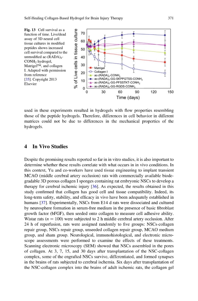
Fig. 13 Cell survival as a
function of time. Live/dead
assay of 3D neural cell
tissue cultures in modified
peptides shows increased
cell survival compared to the
unmodified ac-(RADA) 4 -
CONH 2 hydrogel,
Matrigel™, and collagen
I. Adapted with permission
from reference
[35]. Copyright 2013
Elsevier
Self-Healing Collagen-Based Hydrogel for Brain Injury Therapy
371
those of the peptide hydrogels. Therefore, differences in cell behavior in different
matrices could not be due to differences in the mechanical properties of the
hydrogels.
4 In Vivo Studies
Despite the promising results reported so far in in vitro studies, it is also important to
determine whether these results correlate with what occurs in in vivo conditions. In
this context, Yu and co-workers have used tissue engineering to implant transient
MCAO (middle cerebral artery occlusion) rats with commercially available biodegradable 3D porous collagen I sponges containing rat embryonic NSCs to develop a
therapy for cerebral ischemic injury [36]. As expected, the results obtained in this
study confirmed that collagen has good cell and tissue compatibility. Indeed, its
long-term safety, stability, and efficacy in vivo have been adequately established in
humans [37]. Experimentally, NSCs from E14 d rats were dissociated and cultured
by neurosphere formation in serum-free medium in the presence of basic fibroblast
growth factor (bFGF), then seeded onto collagen to measure cell adhesive ability.
Wistar rats (n ¼ 100) were subjected to 2 h middle cerebral artery occlusion. After
24 h of reperfusion, rats were assigned randomly to five groups: NSCs-collagen
repair group, NSCs repair group, unseeded collagen repair group, MCAO medium
group, and sham group. Neurological, immunohistological, and electronic microscope assessments were performed to examine the effects of these treatments.
Scanning electronic microscopy (SEM) showed that NSCs assembled in the pores
of collagen. At 3, 7, 15, and 30 days after transplantation of the NSC-collagen
complex, some of the engrafted NSCs survive, differentiated, and formed synapses
in the brains of rats subjected to cerebral ischemia. Six days after transplantation of
the NSC-collagen complex into the brains of adult ischemic rats, the collagen gel
Fig. 13 Cell survival as a
function of time. Live/dead
assay of 3D neural cell
tissue cultures in modified
peptides shows increased
cell survival compared to the
unmodified ac-(RADA) 4 -
CONH 2 hydrogel,
Matrigel™, and collagen
I. Adapted with permission
from reference
[35]. Copyright 2013
Elsevier
Self-Healing Collagen-Based Hydrogel for Brain Injury Therapy
371