

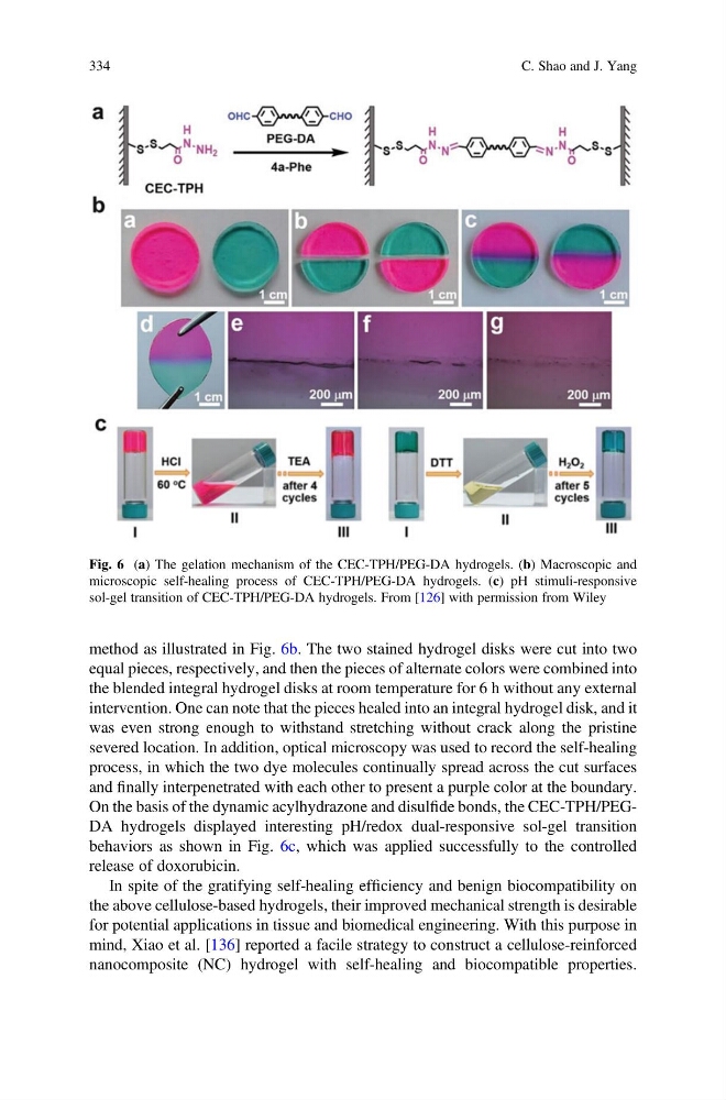
method as illustrated in Fig. 6b. The two stained hydrogel disks were cut into two
equal pieces, respectively, and then the pieces of alternate colors were combined into
the blended integral hydrogel disks at room temperature for 6 h without any external
intervention. One can note that the pieces healed into an integral hydrogel disk, and it
was even strong enough to withstand stretching without crack along the pristine
severed location. In addition, optical microscopy was used to record the self-healing
process, in which the two dye molecules continually spread across the cut surfaces
and finally interpenetrated with each other to present a purple color at the boundary.
On the basis of the dynamic acylhydrazone and disulfide bonds, the CEC-TPH/PEGDA hydrogels displayed interesting pH/redox dual-responsive sol-gel transition
behaviors as shown in Fig. 6c, which was applied successfully to the controlled
release of doxorubicin.
In spite of the gratifying self-healing efficiency and benign biocompatibility on
the above cellulose-based hydrogels, their improved mechanical strength is desirable
for potential applications in tissue and biomedical engineering. With this purpose in
mind, Xiao et al. [136] reported a facile strategy to construct a cellulose-reinforced
nanocomposite (NC) hydrogel with self-healing and biocompatible properties.
Fig. 6 (a) The gelation mechanism of the CEC-TPH/PEG-DA hydrogels. (b) Macroscopic and
microscopic self-healing process of CEC-TPH/PEG-DA hydrogels. (c) pH stimuli-responsive
sol-gel transition of CEC-TPH/PEG-DA hydrogels. From [126] with permission from Wiley
334
C. Shao and J. Yang
equal pieces, respectively, and then the pieces of alternate colors were combined into
the blended integral hydrogel disks at room temperature for 6 h without any external
intervention. One can note that the pieces healed into an integral hydrogel disk, and it
was even strong enough to withstand stretching without crack along the pristine
severed location. In addition, optical microscopy was used to record the self-healing
process, in which the two dye molecules continually spread across the cut surfaces
and finally interpenetrated with each other to present a purple color at the boundary.
On the basis of the dynamic acylhydrazone and disulfide bonds, the CEC-TPH/PEGDA hydrogels displayed interesting pH/redox dual-responsive sol-gel transition
behaviors as shown in Fig. 6c, which was applied successfully to the controlled
release of doxorubicin.
In spite of the gratifying self-healing efficiency and benign biocompatibility on
the above cellulose-based hydrogels, their improved mechanical strength is desirable
for potential applications in tissue and biomedical engineering. With this purpose in
mind, Xiao et al. [136] reported a facile strategy to construct a cellulose-reinforced
nanocomposite (NC) hydrogel with self-healing and biocompatible properties.
Fig. 6 (a) The gelation mechanism of the CEC-TPH/PEG-DA hydrogels. (b) Macroscopic and
microscopic self-healing process of CEC-TPH/PEG-DA hydrogels. (c) pH stimuli-responsive
sol-gel transition of CEC-TPH/PEG-DA hydrogels. From [126] with permission from Wiley
334
C. Shao and J. Yang