

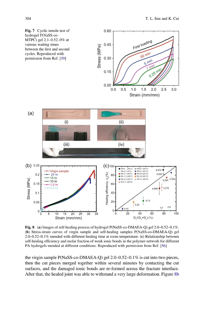
the virgin sample P(NaSS-co-DMAEA-Q) gel 2.0–0.52–0.1% is cut into two pieces,
then the cut pieces merged together within several minutes by contacting the cut
surfaces, and the damaged ionic bonds are re-formed across the fracture interface.
After that, the healed joint was able to withstand a very large deformation. Figure 8b
0.0 0.5 1.0 1.5 2.0 2.5 3.0
0.00
0.15
0.30
0.45
0.60
0 . 2 5 m in
1 m in
5 m in
3 0 m in
Stress (MPa)
Strain (mm/mm)
F ir s t lo a d in g
Fig. 7 Cyclic tensile test of
hydrogel P(NaSS-coMTPC) gel 2.1–0.52–0% at
various waiting times
between the first and second
cycles. Reproduced with
permission from Ref. [39]
0
0.05
0.1
0.15
0.2
0.25
0
5
10 15 20 25 30 35
Virgin sample
24 hr
15 hr
10 hr
1.0 hr
0.5 hr
)
a
P
M
(
s
s
e
r
t
S
Strain (mm/mm)
(b)
(a)
(c)
0
2 0
4 0
6 0
8 0
1 0 0
0
20
40
60
80
100
0.075
1.8
0.036
0.062
2.8
0.40
0.23
0.13
0.085
0.083
PA-0 (25°C)
PA-0.1 (25°C)
PA-0.1 (4°C)
PA-0.1 (37°C)
PA-0.1 (50°C)
PA-0.3 (25°C)
PA-1.0 (25°C)
PA-2.0 (25°C)
PA-4.0 (25°C)
PA ∗ -0 (25°C)
PA ∗ -0 (55°C)
,
h
w
(%
y
c
n
e
i
c
i
f
f
e
g
n
i
l
a
e
H
)
G v /(G e +G v )
0.072
Fig. 8 (a) Images of self-healing process of hydrogel P(NaSS-co-DMAEA-Q) gel 2.0–0.52–0.1%.
(b) Stress-strain curves of virgin sample and self-healing samples P(NaSS-co-DMAEA-Q) gel
2.0–0.52–0.1% mended with different healing time at room temperature. (c) Relationship between
self-healing efficiency and molar fraction of weak ionic bonds in the polymer network for different
PA hydrogels mended at different conditions. Reproduced with permission from Ref. [56]
304
T. L. Sun and K. Cui
then the cut pieces merged together within several minutes by contacting the cut
surfaces, and the damaged ionic bonds are re-formed across the fracture interface.
After that, the healed joint was able to withstand a very large deformation. Figure 8b
0.0 0.5 1.0 1.5 2.0 2.5 3.0
0.00
0.15
0.30
0.45
0.60
0 . 2 5 m in
1 m in
5 m in
3 0 m in
Stress (MPa)
Strain (mm/mm)
F ir s t lo a d in g
Fig. 7 Cyclic tensile test of
hydrogel P(NaSS-coMTPC) gel 2.1–0.52–0% at
various waiting times
between the first and second
cycles. Reproduced with
permission from Ref. [39]
0
0.05
0.1
0.15
0.2
0.25
0
5
10 15 20 25 30 35
Virgin sample
24 hr
15 hr
10 hr
1.0 hr
0.5 hr
)
a
P
M
(
s
s
e
r
t
S
Strain (mm/mm)
(b)
(a)
(c)
0
2 0
4 0
6 0
8 0
1 0 0
0
20
40
60
80
100
0.075
1.8
0.036
0.062
2.8
0.40
0.23
0.13
0.085
0.083
PA-0 (25°C)
PA-0.1 (25°C)
PA-0.1 (4°C)
PA-0.1 (37°C)
PA-0.1 (50°C)
PA-0.3 (25°C)
PA-1.0 (25°C)
PA-2.0 (25°C)
PA-4.0 (25°C)
PA ∗ -0 (25°C)
PA ∗ -0 (55°C)
,
h
w
(%
y
c
n
e
i
c
i
f
f
e
g
n
i
l
a
e
H
)
G v /(G e +G v )
0.072
Fig. 8 (a) Images of self-healing process of hydrogel P(NaSS-co-DMAEA-Q) gel 2.0–0.52–0.1%.
(b) Stress-strain curves of virgin sample and self-healing samples P(NaSS-co-DMAEA-Q) gel
2.0–0.52–0.1% mended with different healing time at room temperature. (c) Relationship between
self-healing efficiency and molar fraction of weak ionic bonds in the polymer network for different
PA hydrogels mended at different conditions. Reproduced with permission from Ref. [56]
304
T. L. Sun and K. Cui