

2.3 pMMO
39
represents a promising avenue for the establishment of catalytic processes for the
conversion of methane to useful chemicals, their protein structure and enzymatic
properties are currently being investigated. At present, the changes in the protein
structure, the structure of the catalytic site, and the reaction mechanism of pMMO
are much less well understood than those of sMMO.
2.3.1 Overall Protein Structure
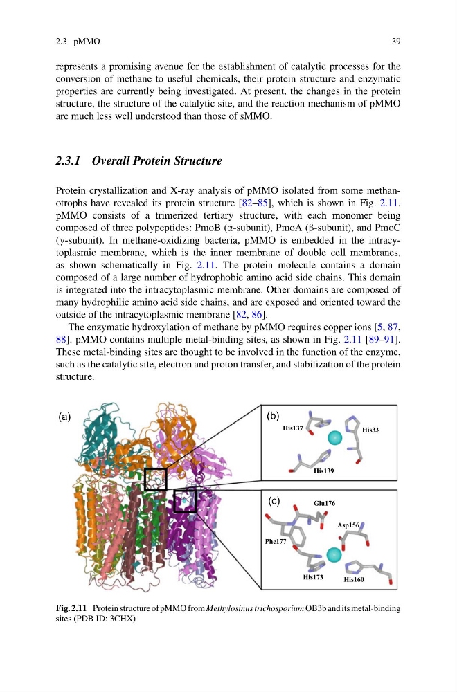
Protein crystallization and X-ray analysis of pMMO isolated from some methanotrophs have revealed its protein structure [82–85], which is shown in Fig. 2.11.
pMMO consists of a trimerized tertiary structure, with each monomer being
composed of three polypeptides: PmoB (α-subunit), PmoA (β-subunit), and PmoC
(γ-subunit). In methane-oxidizing bacteria, pMMO is embedded in the intracytoplasmic membrane, which is the inner membrane of double cell membranes,
as shown schematically in Fig. 2.11. The protein molecule contains a domain
composed of a large number of hydrophobic amino acid side chains. This domain
is integrated into the intracytoplasmic membrane. Other domains are composed of
many hydrophilic amino acid side chains, and are exposed and oriented toward the
outside of the intracytoplasmic membrane [82, 86].
The enzymatic hydroxylation of methane by pMMO requires copper ions [5, 87,
88]. pMMO contains multiple metal-binding sites, as shown in Fig. 2.11 [89–91].
These metal-binding sites are thought to be involved in the function of the enzyme,
such as the catalytic site, electron and proton transfer, and stabilization of the protein
structure.
(a)
(b)
(c)
His33
His137
His139
Glu176
Phe177
His173
His160
Asp156
Fig. 2.11 Protein structure of pMMO from Methylosinus trichosporium OB3b and its metal-binding
sites (PDB ID: 3CHX)
39
represents a promising avenue for the establishment of catalytic processes for the
conversion of methane to useful chemicals, their protein structure and enzymatic
properties are currently being investigated. At present, the changes in the protein
structure, the structure of the catalytic site, and the reaction mechanism of pMMO
are much less well understood than those of sMMO.
2.3.1 Overall Protein Structure
Protein crystallization and X-ray analysis of pMMO isolated from some methanotrophs have revealed its protein structure [82–85], which is shown in Fig. 2.11.
pMMO consists of a trimerized tertiary structure, with each monomer being
composed of three polypeptides: PmoB (α-subunit), PmoA (β-subunit), and PmoC
(γ-subunit). In methane-oxidizing bacteria, pMMO is embedded in the intracytoplasmic membrane, which is the inner membrane of double cell membranes,
as shown schematically in Fig. 2.11. The protein molecule contains a domain
composed of a large number of hydrophobic amino acid side chains. This domain
is integrated into the intracytoplasmic membrane. Other domains are composed of
many hydrophilic amino acid side chains, and are exposed and oriented toward the
outside of the intracytoplasmic membrane [82, 86].
The enzymatic hydroxylation of methane by pMMO requires copper ions [5, 87,
88]. pMMO contains multiple metal-binding sites, as shown in Fig. 2.11 [89–91].
These metal-binding sites are thought to be involved in the function of the enzyme,
such as the catalytic site, electron and proton transfer, and stabilization of the protein
structure.
(a)
(b)
(c)
His33
His137
His139
Glu176
Phe177
His173
His160
Asp156
Fig. 2.11 Protein structure of pMMO from Methylosinus trichosporium OB3b and its metal-binding
sites (PDB ID: 3CHX)