

26
2 Selective Production of Methanol …
MMOH. When the MMOB unit was removed, the turnover number decreased 150fold, and the initial reaction rate with oxygen decreased 1000-fold compared to when
this protein was present [12].
2.2.2 Coordination Structure of the Di-Nuclear Iron Cluster
Crystallographic studies of MMOH indicate that only one iron-binding site is present
in the MMOH monomer, and that two iron ions are bound at this site [10, 13–15].
The two iron ions are bridged by a carboxylate group and an oxygen. As mentioned
above, this di-nuclear iron site is the catalytic center for the oxidation of methane to
methanol.
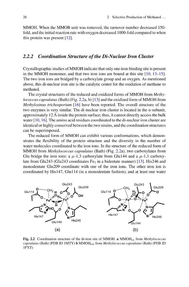
The crystal structures of the reduced and oxidized forms of MMOH from Methylococcus capsulatus (Bath) (Fig. 2.2a, b) [13] and the oxidized form of MMOH from
Methylosinus trichosporium [16] have been reported. The overall structure of the
two enzymes is very similar. The di-nuclear iron cluster is located in the α-subunit,
approximately 12 Å inside the protein surface; thus, it cannot directly access the bulk
water [10, 16]. The amino acid residues coordinated to the di-nuclear iron cluster are
identical or highly conserved between the two strains, and the coordination structures
can be superimposed.
The reduced form of MMOH can exhibit various conformations, which demonstrates the flexibility of the protein structure and the diversity in the number of
water molecules coordinated to the iron ions. In the structure of the reduced form of
MMOH from Methylococcus capsulatus (Bath) (Fig. 2.2a), two carboxylates from
Glu bridge the iron ions: a μ-1,3 carboxylate from Glu144 and a μ-1,1 carboxylate from Glu243 (Glu243 coordinates Fe 2 in a bidentate manner) [13]. His246 and
monodentate Glu209 coordinate with one of the iron ions. The other iron ion is
coordinated by His147, Glu114 (in a monodentate fashion), and at least one water
Fe
II
Fe
II
O
H 2
O
O
O
N
N
N
N
O
O
O
O
O
O
H 2
Glu209
Glu243
His246
His147
Glu144
Glu114
Glu209
Glu243
His246
His147
Glu144
Glu114
Fe
II
Fe
II
O
O
H
H
O
HO O
N
N
N
N
OH
O
O
O
O
O
H 2
(a)
(b)
Fig. 2.2 Coordination structure of the di-iron site of MMOH. a MMOH ox from Methylococcus
capsulatus (Bath) (PDB ID 1MTY) b MMOH red from Methylococcus capsulatus (Bath) (PDB ID
1FYZ)
2 Selective Production of Methanol …
MMOH. When the MMOB unit was removed, the turnover number decreased 150fold, and the initial reaction rate with oxygen decreased 1000-fold compared to when
this protein was present [12].
2.2.2 Coordination Structure of the Di-Nuclear Iron Cluster
Crystallographic studies of MMOH indicate that only one iron-binding site is present
in the MMOH monomer, and that two iron ions are bound at this site [10, 13–15].
The two iron ions are bridged by a carboxylate group and an oxygen. As mentioned
above, this di-nuclear iron site is the catalytic center for the oxidation of methane to
methanol.
The crystal structures of the reduced and oxidized forms of MMOH from Methylococcus capsulatus (Bath) (Fig. 2.2a, b) [13] and the oxidized form of MMOH from
Methylosinus trichosporium [16] have been reported. The overall structure of the
two enzymes is very similar. The di-nuclear iron cluster is located in the α-subunit,
approximately 12 Å inside the protein surface; thus, it cannot directly access the bulk
water [10, 16]. The amino acid residues coordinated to the di-nuclear iron cluster are
identical or highly conserved between the two strains, and the coordination structures
can be superimposed.
The reduced form of MMOH can exhibit various conformations, which demonstrates the flexibility of the protein structure and the diversity in the number of
water molecules coordinated to the iron ions. In the structure of the reduced form of
MMOH from Methylococcus capsulatus (Bath) (Fig. 2.2a), two carboxylates from
Glu bridge the iron ions: a μ-1,3 carboxylate from Glu144 and a μ-1,1 carboxylate from Glu243 (Glu243 coordinates Fe 2 in a bidentate manner) [13]. His246 and
monodentate Glu209 coordinate with one of the iron ions. The other iron ion is
coordinated by His147, Glu114 (in a monodentate fashion), and at least one water
Fe
II
Fe
II
O
H 2
O
O
O
N
N
N
N
O
O
O
O
O
O
H 2
Glu209
Glu243
His246
His147
Glu144
Glu114
Glu209
Glu243
His246
His147
Glu144
Glu114
Fe
II
Fe
II
O
O
H
H
O
HO O
N
N
N
N
OH
O
O
O
O
O
H 2
(a)
(b)
Fig. 2.2 Coordination structure of the di-iron site of MMOH. a MMOH ox from Methylococcus
capsulatus (Bath) (PDB ID 1MTY) b MMOH red from Methylococcus capsulatus (Bath) (PDB ID
1FYZ)