

50
S. Kerbstadt et al.
3.2.2 Photoelectron Tomography
Coherent control of ultrafast dynamics using polarization-shaped pulses exploits the
vectorial character of light-matter interactions. This opens the door to the manipulation of spatial aspects of quantum phenomena such as asymmetries in the wave
function of a quantum system or the directionality of quantum processes. In order
to utilize the full potential of spatial control, the application of 3D detection techniques providing vectorial information on the dynamics is crucial as well. Among the
various detection schemes, based on the measurement of either photons or massive
particles, the measurement of photoelectrons is particularly attractive due to its high
detection efficiencies and the wealth of information provided by the 3D PMD. Different methods have been devised to measure the PMD, yielding highly differential
information on the processes under investigation. For example, fundamental aspects
of light-matter interactions have been studied using cold target recoil ion momentum
spectroscopy (COLTRIMS) [10]. The COLTRIMS technique permits the reconstruction the 3D PMD by coincidence measurement of photoelectrons and photoions with
angular resolution. Another well-established method to measure the photoelectron
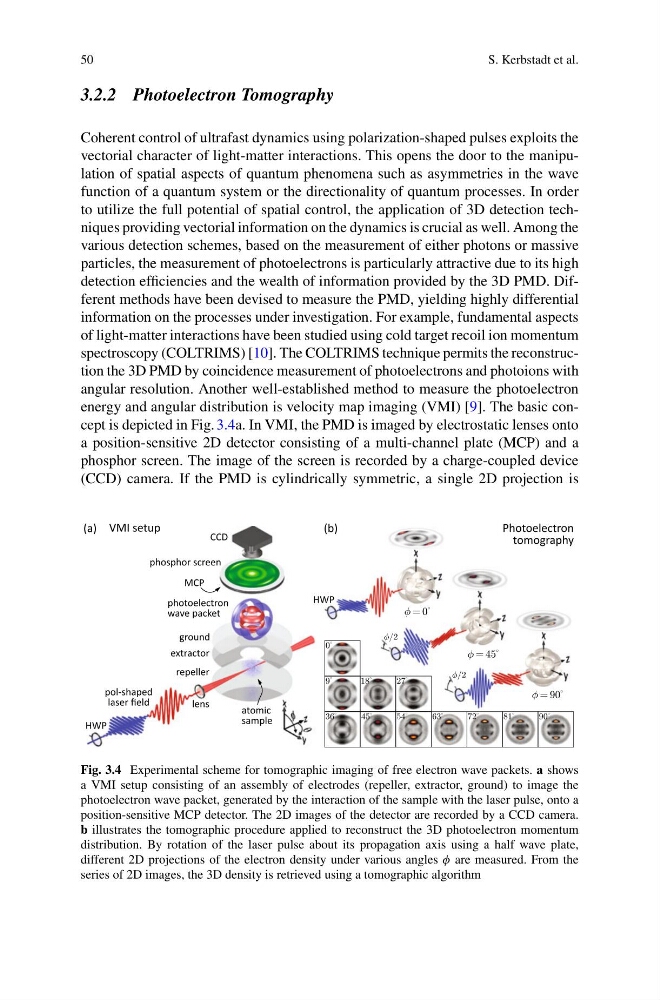
energy and angular distribution is velocity map imaging (VMI) [9]. The basic concept is depicted in Fig. 3.4a. In VMI, the PMD is imaged by electrostatic lenses onto
a position-sensitive 2D detector consisting of a multi-channel plate (MCP) and a
phosphor screen. The image of the screen is recorded by a charge-coupled device
(CCD) camera. If the PMD is cylindrically symmetric, a single 2D projection is
Fig. 3.4 Experimental scheme for tomographic imaging of free electron wave packets. a shows
a VMI setup consisting of an assembly of electrodes (repeller, extractor, ground) to image the
photoelectron wave packet, generated by the interaction of the sample with the laser pulse, onto a
position-sensitive MCP detector. The 2D images of the detector are recorded by a CCD camera.
b illustrates the tomographic procedure applied to reconstruct the 3D photoelectron momentum
distribution. By rotation of the laser pulse about its propagation axis using a half wave plate,
different 2D projections of the electron density under various angles φ are measured. From the
series of 2D images, the 3D density is retrieved using a tomographic algorithm
S. Kerbstadt et al.
3.2.2 Photoelectron Tomography
Coherent control of ultrafast dynamics using polarization-shaped pulses exploits the
vectorial character of light-matter interactions. This opens the door to the manipulation of spatial aspects of quantum phenomena such as asymmetries in the wave
function of a quantum system or the directionality of quantum processes. In order
to utilize the full potential of spatial control, the application of 3D detection techniques providing vectorial information on the dynamics is crucial as well. Among the
various detection schemes, based on the measurement of either photons or massive
particles, the measurement of photoelectrons is particularly attractive due to its high
detection efficiencies and the wealth of information provided by the 3D PMD. Different methods have been devised to measure the PMD, yielding highly differential
information on the processes under investigation. For example, fundamental aspects
of light-matter interactions have been studied using cold target recoil ion momentum
spectroscopy (COLTRIMS) [10]. The COLTRIMS technique permits the reconstruction the 3D PMD by coincidence measurement of photoelectrons and photoions with
angular resolution. Another well-established method to measure the photoelectron
energy and angular distribution is velocity map imaging (VMI) [9]. The basic concept is depicted in Fig. 3.4a. In VMI, the PMD is imaged by electrostatic lenses onto
a position-sensitive 2D detector consisting of a multi-channel plate (MCP) and a
phosphor screen. The image of the screen is recorded by a charge-coupled device
(CCD) camera. If the PMD is cylindrically symmetric, a single 2D projection is
Fig. 3.4 Experimental scheme for tomographic imaging of free electron wave packets. a shows
a VMI setup consisting of an assembly of electrodes (repeller, extractor, ground) to image the
photoelectron wave packet, generated by the interaction of the sample with the laser pulse, onto a
position-sensitive MCP detector. The 2D images of the detector are recorded by a CCD camera.
b illustrates the tomographic procedure applied to reconstruct the 3D photoelectron momentum
distribution. By rotation of the laser pulse about its propagation axis using a half wave plate,
different 2D projections of the electron density under various angles φ are measured. From the
series of 2D images, the 3D density is retrieved using a tomographic algorithm