

6.4 Complex Methodology for Reverse Engineering …
569
Digitalization workplace is fitted with a motorized optical microscope working
in visible and UV ranges, with a specialized color digital camera with the possibility
of performing micro-photography in visible and UV ranges, specialized software for
microscopy, and a precision motorized scanning object table.
Workplace controlled by the PC (workstation) supports:
– Fully automated (motorized) condenser for work with lenses from 5x to 150x in
the modes of light field, dark field and phase contrast;
– Fully automated fluorescence system with a motorized fluorescent filter system
in a cubic holder with a turret;
– Automatic aperture selection when changing lenses and filters;
– Automatic measurement of transmitted and incident radiation intensity in case of
change of lenses and filters;
– Automatic monitoring of the stability of color temperature of transmitted and
incident radiation;
– Automatic saving of all microscope and camera settings for current image;
– Automatic saving of all microscope and camera settings for further work;
– Motorized Z-drive for automatic parfocality adjustment.
Specialized software of the microscopy complex allowed for automated digitalization of the entire chip after performed initial settings and adjustments.
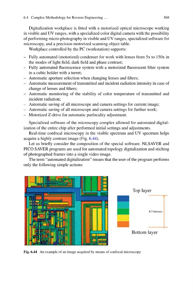
Real-time confocal microscopy in the visible spectrum and UV spectrum helps
acquire a highly contrast image (Fig. 6.44).
Let us briefly consider the composition of the special software. NLSAVER and
PICO SAVER programs are used for automated topology digitalization and stiching
of photographed frames into a single video image.
The term “automated digitalization” means that the user of the program performs
only the following simple actions:
Fig. 6.44 An example of an image acquired by means of confocal microscopy
569
Digitalization workplace is fitted with a motorized optical microscope working
in visible and UV ranges, with a specialized color digital camera with the possibility
of performing micro-photography in visible and UV ranges, specialized software for
microscopy, and a precision motorized scanning object table.
Workplace controlled by the PC (workstation) supports:
– Fully automated (motorized) condenser for work with lenses from 5x to 150x in
the modes of light field, dark field and phase contrast;
– Fully automated fluorescence system with a motorized fluorescent filter system
in a cubic holder with a turret;
– Automatic aperture selection when changing lenses and filters;
– Automatic measurement of transmitted and incident radiation intensity in case of
change of lenses and filters;
– Automatic monitoring of the stability of color temperature of transmitted and
incident radiation;
– Automatic saving of all microscope and camera settings for current image;
– Automatic saving of all microscope and camera settings for further work;
– Motorized Z-drive for automatic parfocality adjustment.
Specialized software of the microscopy complex allowed for automated digitalization of the entire chip after performed initial settings and adjustments.
Real-time confocal microscopy in the visible spectrum and UV spectrum helps
acquire a highly contrast image (Fig. 6.44).
Let us briefly consider the composition of the special software. NLSAVER and
PICO SAVER programs are used for automated topology digitalization and stiching
of photographed frames into a single video image.
The term “automated digitalization” means that the user of the program performs
only the following simple actions:
Fig. 6.44 An example of an image acquired by means of confocal microscopy