

178
V. Laxmi et al.
six shades of red, where each shade represents the corresponding hemoglobin. This
device is capable of identifying anemia and its severity but is not capable of quantifying the result [51]. Although many methods have been reported for hemoglobin
measurement, only a few provide accurate results. The HiCN is considered to be the
gold standard method; however, it is time consuming, requires a spectrophotometer,
and is unsafe due to the use of cyanide [45, 52]. The automated analyzers are accurate but they require a lab infrastructure and are expensive. The requirement for a
portable, efficient, and low-cost microfluidic-based device therefore still exists.
Clinical methods to measure hematocrit are by means of centrifugation, the CBC
(impedance), conductivity, or by indirect method using the hemoglobin value [41].
The reference method is the centrifugation technique. In this method, blood is drawn
into a capillary and rotated at high speeds using a centrifuge and the hematocrit
(Hct) can be directly read from the scale (the ratio of RBCs to the total volume).
Most commonly, the automated analyzers (based on Coulter impedance principle)
are used, and hematocrit is measured from average size and number of RBCs. Hct
can also be measured using a Point-of-care device, such as i-STAT blood gas analyzer
[41]. These analyzers measure Hct using the principle of electrical conductivity or
by evaluating Hct from the hemoglobin value. In recent work, Berry et al. [53]
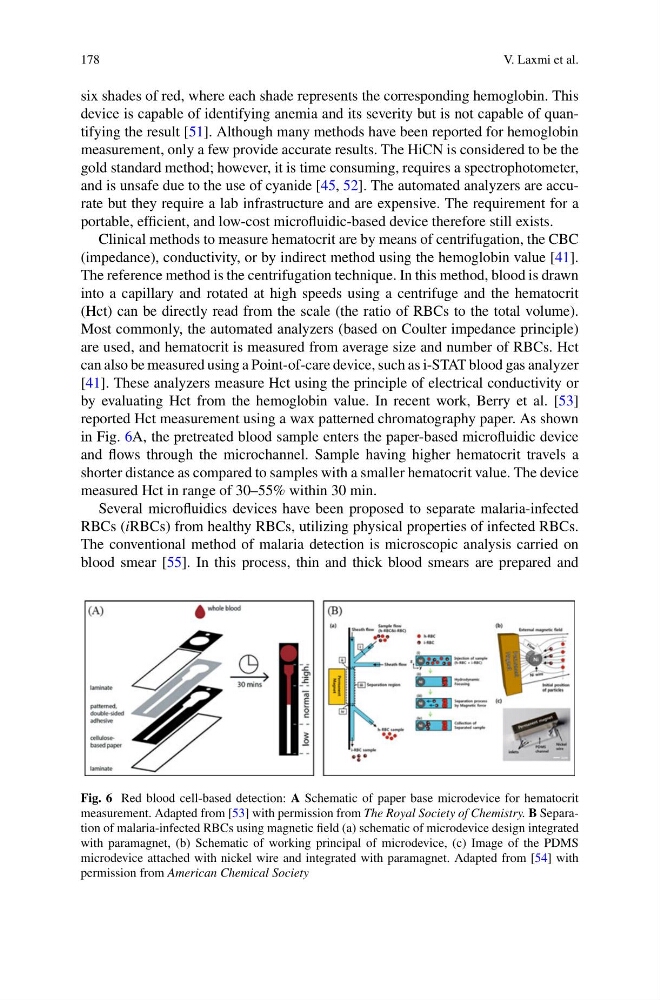
reported Hct measurement using a wax patterned chromatography paper. As shown
in Fig. 6A, the pretreated blood sample enters the paper-based microfluidic device
and flows through the microchannel. Sample having higher hematocrit travels a
shorter distance as compared to samples with a smaller hematocrit value. The device
measured Hct in range of 30–55% within 30 min.
Several microfluidics devices have been proposed to separate malaria-infected
RBCs (iRBCs) from healthy RBCs, utilizing physical properties of infected RBCs.
The conventional method of malaria detection is microscopic analysis carried on
blood smear [55]. In this process, thin and thick blood smears are prepared and
Fig. 6 Red blood cell-based detection: A Schematic of paper base microdevice for hematocrit
measurement. Adapted from [53] with permission from The Royal Society of Chemistry. B Separation of malaria-infected RBCs using magnetic field (a) schematic of microdevice design integrated
with paramagnet, (b) Schematic of working principal of microdevice, (c) Image of the PDMS
microdevice attached with nickel wire and integrated with paramagnet. Adapted from [54] with
permission from American Chemical Society
V. Laxmi et al.
six shades of red, where each shade represents the corresponding hemoglobin. This
device is capable of identifying anemia and its severity but is not capable of quantifying the result [51]. Although many methods have been reported for hemoglobin
measurement, only a few provide accurate results. The HiCN is considered to be the
gold standard method; however, it is time consuming, requires a spectrophotometer,
and is unsafe due to the use of cyanide [45, 52]. The automated analyzers are accurate but they require a lab infrastructure and are expensive. The requirement for a
portable, efficient, and low-cost microfluidic-based device therefore still exists.
Clinical methods to measure hematocrit are by means of centrifugation, the CBC
(impedance), conductivity, or by indirect method using the hemoglobin value [41].
The reference method is the centrifugation technique. In this method, blood is drawn
into a capillary and rotated at high speeds using a centrifuge and the hematocrit
(Hct) can be directly read from the scale (the ratio of RBCs to the total volume).
Most commonly, the automated analyzers (based on Coulter impedance principle)
are used, and hematocrit is measured from average size and number of RBCs. Hct
can also be measured using a Point-of-care device, such as i-STAT blood gas analyzer
[41]. These analyzers measure Hct using the principle of electrical conductivity or
by evaluating Hct from the hemoglobin value. In recent work, Berry et al. [53]
reported Hct measurement using a wax patterned chromatography paper. As shown
in Fig. 6A, the pretreated blood sample enters the paper-based microfluidic device
and flows through the microchannel. Sample having higher hematocrit travels a
shorter distance as compared to samples with a smaller hematocrit value. The device
measured Hct in range of 30–55% within 30 min.
Several microfluidics devices have been proposed to separate malaria-infected
RBCs (iRBCs) from healthy RBCs, utilizing physical properties of infected RBCs.
The conventional method of malaria detection is microscopic analysis carried on
blood smear [55]. In this process, thin and thick blood smears are prepared and
Fig. 6 Red blood cell-based detection: A Schematic of paper base microdevice for hematocrit
measurement. Adapted from [53] with permission from The Royal Society of Chemistry. B Separation of malaria-infected RBCs using magnetic field (a) schematic of microdevice design integrated
with paramagnet, (b) Schematic of working principal of microdevice, (c) Image of the PDMS
microdevice attached with nickel wire and integrated with paramagnet. Adapted from [54] with
permission from American Chemical Society