

20 Twenty-Five Years’ History, Mechanism, and Generality …
425
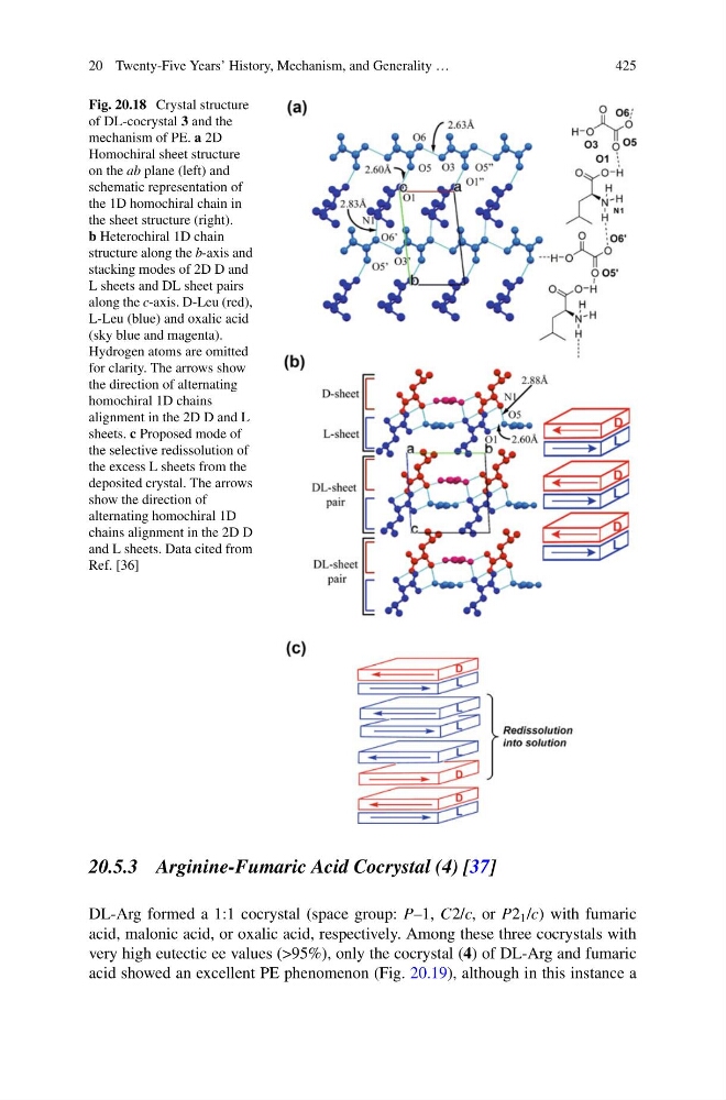
Fig. 20.18 Crystal structure
of DL-cocrystal 3 and the
mechanism of PE. a 2D
Homochiral sheet structure
on the ab plane (left) and
schematic representation of
the 1D homochiral chain in
the sheet structure (right).
b Heterochiral 1D chain
structure along the b-axis and
stacking modes of 2D D and
L sheets and DL sheet pairs
along the c-axis. D-Leu (red),
L-Leu (blue) and oxalic acid
(sky blue and magenta).
Hydrogen atoms are omitted
for clarity. The arrows show
the direction of alternating
homochiral 1D chains
alignment in the 2D D and L
sheets. c Proposed mode of
the selective redissolution of
the excess L sheets from the
deposited crystal. The arrows
show the direction of
alternating homochiral 1D
chains alignment in the 2D D
and L sheets. Data cited from
Ref. [36]
20.5.3 Arginine-Fumaric Acid Cocrystal (4) [37]
DL-Arg formed a 1:1 cocrystal (space group: P–1, C2/c, or P2 1 /c) with fumaric
acid, malonic acid, or oxalic acid, respectively. Among these three cocrystals with
very high eutectic ee values (>95%), only the cocrystal (4) of DL-Arg and fumaric
acid showed an excellent PE phenomenon (Fig. 20.19), although in this instance a
425
Fig. 20.18 Crystal structure
of DL-cocrystal 3 and the
mechanism of PE. a 2D
Homochiral sheet structure
on the ab plane (left) and
schematic representation of
the 1D homochiral chain in
the sheet structure (right).
b Heterochiral 1D chain
structure along the b-axis and
stacking modes of 2D D and
L sheets and DL sheet pairs
along the c-axis. D-Leu (red),
L-Leu (blue) and oxalic acid
(sky blue and magenta).
Hydrogen atoms are omitted
for clarity. The arrows show
the direction of alternating
homochiral 1D chains
alignment in the 2D D and L
sheets. c Proposed mode of
the selective redissolution of
the excess L sheets from the
deposited crystal. The arrows
show the direction of
alternating homochiral 1D
chains alignment in the 2D D
and L sheets. Data cited from
Ref. [36]
20.5.3 Arginine-Fumaric Acid Cocrystal (4) [37]
DL-Arg formed a 1:1 cocrystal (space group: P–1, C2/c, or P2 1 /c) with fumaric
acid, malonic acid, or oxalic acid, respectively. Among these three cocrystals with
very high eutectic ee values (>95%), only the cocrystal (4) of DL-Arg and fumaric
acid showed an excellent PE phenomenon (Fig. 20.19), although in this instance a