

14 Luminescent Crystal–Control of Excited-State …
283
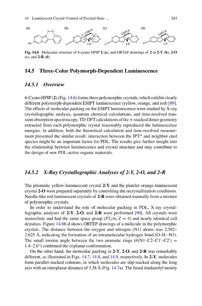
Fig. 14.6 Molecular structure of 6-cyano HPIP 2 (a), and ORTEP drawings of 2 in 2-Y (b), 2-O
(c), and 2-R (d)
14.5 Three-Color Polymorph-Dependent Luminescence
14.5.1 Overview
6-Cyano HPIP (2) (Fig. 14.6) forms three polymorphic crystals, which exhibit clearly
different polymorph-dependent ESIPT luminescence (yellow, orange, and red) [89].
The effects of molecular packing on the ESIPT luminescence were studied by X-ray
crystallographic analysis, quantum chemical calculations, and time-resolved transient absorption spectroscopy. TD-DFT calculations of the π-stacked dimer geometry
extracted from each polymorphic crystal reasonably reproduced the luminescence
energies. In addition, both the theoretical calculation and time-resolved measurement presented the similar result: interaction between the IPT* and neighbor enol
species might be an important factor for PDL. The results give further insight into
the relationship between luminescence and crystal structure and may contribute to
the design of new PDL-active organic materials.
14.5.2 X-Ray Crystallographic Analyses of 2-Y, 2-O, and 2-R
The prismatic yellow-luminescent crystal 2-Y and the platelet orange-luminescent
crystal 2-O were prepared separately by controlling the recrystallization conditions.
Needle-like red-luminescent crystals of 2-R were obtained manually from a mixture
of polymorphic crystals.
In order to understand the role of molecular packing in PDL, X-ray crystallographic analyses of 2-Y, 2-O, and 2-R were performed [90]. All crystals were
monoclinic and had the same space group (P2 1 /n, Z = 4) and nearly identical cell
densities. Figure 14.6b-d shows ORTEP drawings of a molecule in the polymorphic
crystals. The distance between the oxygen and nitrogen (N1) atoms was 2.582–
2.625 Å, indicating the formation of an intramolecular hydrogen bond (O–H···N1).
The small torsion angle between the two aromatic rings (θ (N1–C2–C1
–C2
) =
1.4–2.6°) confirmed the coplanar conformation.
On the other hand, the molecular packing in 2-Y, 2-O, and 2-R was remarkably
different, as illustrated in Figs. 14.7, 14.8, and 14.9, respectively. In 2-Y, molecules
form parallel-stacked columns, in which molecules are slip-stacked along the long
axis with an interplanar distance of 3.38 Å (Fig. 14.7a). The fused imidazolyl moiety
283
Fig. 14.6 Molecular structure of 6-cyano HPIP 2 (a), and ORTEP drawings of 2 in 2-Y (b), 2-O
(c), and 2-R (d)
14.5 Three-Color Polymorph-Dependent Luminescence
14.5.1 Overview
6-Cyano HPIP (2) (Fig. 14.6) forms three polymorphic crystals, which exhibit clearly
different polymorph-dependent ESIPT luminescence (yellow, orange, and red) [89].
The effects of molecular packing on the ESIPT luminescence were studied by X-ray
crystallographic analysis, quantum chemical calculations, and time-resolved transient absorption spectroscopy. TD-DFT calculations of the π-stacked dimer geometry
extracted from each polymorphic crystal reasonably reproduced the luminescence
energies. In addition, both the theoretical calculation and time-resolved measurement presented the similar result: interaction between the IPT* and neighbor enol
species might be an important factor for PDL. The results give further insight into
the relationship between luminescence and crystal structure and may contribute to
the design of new PDL-active organic materials.
14.5.2 X-Ray Crystallographic Analyses of 2-Y, 2-O, and 2-R
The prismatic yellow-luminescent crystal 2-Y and the platelet orange-luminescent
crystal 2-O were prepared separately by controlling the recrystallization conditions.
Needle-like red-luminescent crystals of 2-R were obtained manually from a mixture
of polymorphic crystals.
In order to understand the role of molecular packing in PDL, X-ray crystallographic analyses of 2-Y, 2-O, and 2-R were performed [90]. All crystals were
monoclinic and had the same space group (P2 1 /n, Z = 4) and nearly identical cell
densities. Figure 14.6b-d shows ORTEP drawings of a molecule in the polymorphic
crystals. The distance between the oxygen and nitrogen (N1) atoms was 2.582–
2.625 Å, indicating the formation of an intramolecular hydrogen bond (O–H···N1).
The small torsion angle between the two aromatic rings (θ (N1–C2–C1
–C2
) =
1.4–2.6°) confirmed the coplanar conformation.
On the other hand, the molecular packing in 2-Y, 2-O, and 2-R was remarkably
different, as illustrated in Figs. 14.7, 14.8, and 14.9, respectively. In 2-Y, molecules
form parallel-stacked columns, in which molecules are slip-stacked along the long
axis with an interplanar distance of 3.38 Å (Fig. 14.7a). The fused imidazolyl moiety