

14 Luminescent Crystal–Control of Excited-State …
277
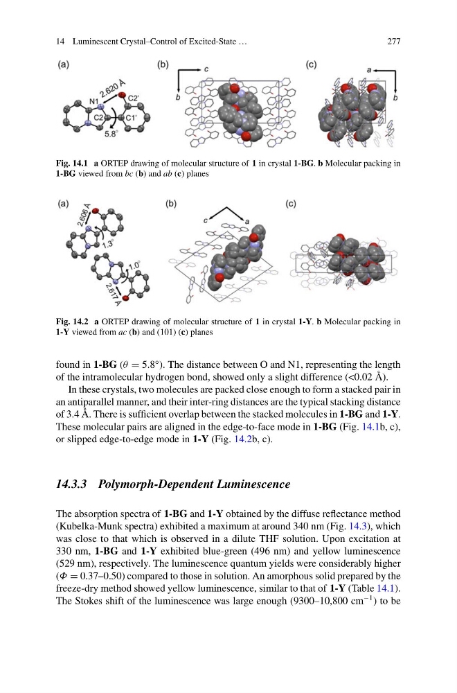
Fig. 14.1 a ORTEP drawing of molecular structure of 1 in crystal 1-BG. b Molecular packing in
1-BG viewed from bc (b) and ab (c) planes
Fig. 14.2 a ORTEP drawing of molecular structure of 1 in crystal 1-Y. b Molecular packing in
1-Y viewed from ac (b) and (101) (c) planes
found in 1-BG (θ = 5.8°). The distance between O and N1, representing the length
of the intramolecular hydrogen bond, showed only a slight difference (<0.02 Å).
In these crystals, two molecules are packed close enough to form a stacked pair in
an antiparallel manner, and their inter-ring distances are the typical stacking distance
of 3.4 Å. There is sufficient overlap between the stacked molecules in 1-BG and 1-Y.
These molecular pairs are aligned in the edge-to-face mode in 1-BG (Fig. 14.1b, c),
or slipped edge-to-edge mode in 1-Y (Fig. 14.2b, c).
14.3.3 Polymorph-Dependent Luminescence
The absorption spectra of 1-BG and 1-Y obtained by the diffuse reflectance method
(Kubelka-Munk spectra) exhibited a maximum at around 340 nm (Fig. 14.3), which
was close to that which is observed in a dilute THF solution. Upon excitation at
330 nm, 1-BG and 1-Y exhibited blue-green (496 nm) and yellow luminescence
(529 nm), respectively. The luminescence quantum yields were considerably higher
(Φ = 0.37–0.50) compared to those in solution. An amorphous solid prepared by the
freeze-dry method showed yellow luminescence, similar to that of 1-Y (Table 14.1).
The Stokes shift of the luminescence was large enough (9300–10,800 cm
−1 ) to be
277
Fig. 14.1 a ORTEP drawing of molecular structure of 1 in crystal 1-BG. b Molecular packing in
1-BG viewed from bc (b) and ab (c) planes
Fig. 14.2 a ORTEP drawing of molecular structure of 1 in crystal 1-Y. b Molecular packing in
1-Y viewed from ac (b) and (101) (c) planes
found in 1-BG (θ = 5.8°). The distance between O and N1, representing the length
of the intramolecular hydrogen bond, showed only a slight difference (<0.02 Å).
In these crystals, two molecules are packed close enough to form a stacked pair in
an antiparallel manner, and their inter-ring distances are the typical stacking distance
of 3.4 Å. There is sufficient overlap between the stacked molecules in 1-BG and 1-Y.
These molecular pairs are aligned in the edge-to-face mode in 1-BG (Fig. 14.1b, c),
or slipped edge-to-edge mode in 1-Y (Fig. 14.2b, c).
14.3.3 Polymorph-Dependent Luminescence
The absorption spectra of 1-BG and 1-Y obtained by the diffuse reflectance method
(Kubelka-Munk spectra) exhibited a maximum at around 340 nm (Fig. 14.3), which
was close to that which is observed in a dilute THF solution. Upon excitation at
330 nm, 1-BG and 1-Y exhibited blue-green (496 nm) and yellow luminescence
(529 nm), respectively. The luminescence quantum yields were considerably higher
(Φ = 0.37–0.50) compared to those in solution. An amorphous solid prepared by the
freeze-dry method showed yellow luminescence, similar to that of 1-Y (Table 14.1).
The Stokes shift of the luminescence was large enough (9300–10,800 cm
−1 ) to be