

396
T. Dewa et al.
TA spectroscopy. Therefore, introducing Cys and chemical modification with A647
did not perturb the environment of the intrinsic chromophores. The absence of CD
signal from A647 implied no excitonic coupling between A647 moieties.
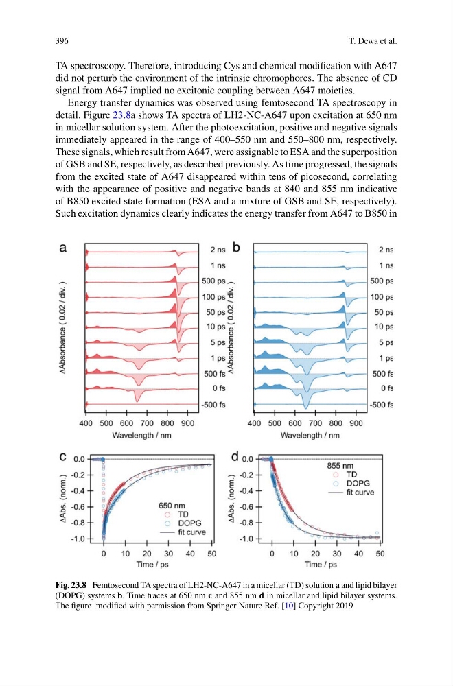
Energy transfer dynamics was observed using femtosecond TA spectroscopy in
detail. Figure 23.8a shows TA spectra of LH2-NC-A647 upon excitation at 650 nm
in micellar solution system. After the photoexcitation, positive and negative signals
immediately appeared in the range of 400–550 nm and 550–800 nm, respectively.
These signals, which result from A647, were assignable to ESA and the superposition
of GSB and SE, respectively, as described previously. As time progressed, the signals
from the excited state of A647 disappeared within tens of picosecond, correlating
with the appearance of positive and negative bands at 840 and 855 nm indicative
of B850 excited state formation (ESA and a mixture of GSB and SE, respectively).
Such excitation dynamics clearly indicates the energy transfer from A647 to B850 in
Fig. 23.8 Femtosecond TA spectra of LH2-NC-A647 in a micellar (TD) solution a and lipid bilayer
(DOPG) systems b. Time traces at 650 nm c and 855 nm d in micellar and lipid bilayer systems.
The figure modified with permission from Springer Nature Ref. [10] Copyright 2019
T. Dewa et al.
TA spectroscopy. Therefore, introducing Cys and chemical modification with A647
did not perturb the environment of the intrinsic chromophores. The absence of CD
signal from A647 implied no excitonic coupling between A647 moieties.
Energy transfer dynamics was observed using femtosecond TA spectroscopy in
detail. Figure 23.8a shows TA spectra of LH2-NC-A647 upon excitation at 650 nm
in micellar solution system. After the photoexcitation, positive and negative signals
immediately appeared in the range of 400–550 nm and 550–800 nm, respectively.
These signals, which result from A647, were assignable to ESA and the superposition
of GSB and SE, respectively, as described previously. As time progressed, the signals
from the excited state of A647 disappeared within tens of picosecond, correlating
with the appearance of positive and negative bands at 840 and 855 nm indicative
of B850 excited state formation (ESA and a mixture of GSB and SE, respectively).
Such excitation dynamics clearly indicates the energy transfer from A647 to B850 in
Fig. 23.8 Femtosecond TA spectra of LH2-NC-A647 in a micellar (TD) solution a and lipid bilayer
(DOPG) systems b. Time traces at 650 nm c and 855 nm d in micellar and lipid bilayer systems.
The figure modified with permission from Springer Nature Ref. [10] Copyright 2019