

19 Photoinduced Morphological Transformation and Photodriven …
341
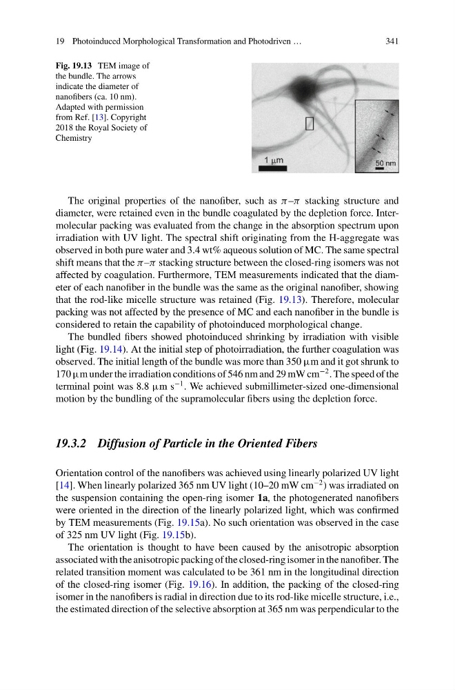
Fig. 19.13 TEM image of
the bundle. The arrows
indicate the diameter of
nanofibers (ca. 10 nm).
Adapted with permission
from Ref. [13]. Copyright
2018 the Royal Society of
Chemistry
The original properties of the nanofiber, such as π –π stacking structure and
diameter, were retained even in the bundle coagulated by the depletion force. Intermolecular packing was evaluated from the change in the absorption spectrum upon
irradiation with UV light. The spectral shift originating from the H-aggregate was
observed in both pure water and 3.4 wt% aqueous solution of MC. The same spectral
shift means that the π –π stacking structure between the closed-ring isomers was not
affected by coagulation. Furthermore, TEM measurements indicated that the diameter of each nanofiber in the bundle was the same as the original nanofiber, showing
that the rod-like micelle structure was retained (Fig. 19.13). Therefore, molecular
packing was not affected by the presence of MC and each nanofiber in the bundle is
considered to retain the capability of photoinduced morphological change.
The bundled fibers showed photoinduced shrinking by irradiation with visible
light (Fig. 19.14). At the initial step of photoirradiation, the further coagulation was
observed. The initial length of the bundle was more than 350 µm and it got shrunk to
170 µm under the irradiation conditions of 546 nm and 29 mW cm
−2 . The speed of the
terminal point was 8.8 µm s
−1 . We achieved submillimeter-sized one-dimensional
motion by the bundling of the supramolecular fibers using the depletion force.
19.3.2 Diffusion of Particle in the Oriented Fibers
Orientation control of the nanofibers was achieved using linearly polarized UV light
[14]. When linearly polarized 365 nm UV light (10–20 mW cm
−2 ) was irradiated on
the suspension containing the open-ring isomer 1a, the photogenerated nanofibers
were oriented in the direction of the linearly polarized light, which was confirmed
by TEM measurements (Fig. 19.15a). No such orientation was observed in the case
of 325 nm UV light (Fig. 19.15b).
The orientation is thought to have been caused by the anisotropic absorption
associated with the anisotropic packing of the closed-ring isomer in the nanofiber. The
related transition moment was calculated to be 361 nm in the longitudinal direction
of the closed-ring isomer (Fig. 19.16). In addition, the packing of the closed-ring
isomer in the nanofibers is radial in direction due to its rod-like micelle structure, i.e.,
the estimated direction of the selective absorption at 365 nm was perpendicular to the
341
Fig. 19.13 TEM image of
the bundle. The arrows
indicate the diameter of
nanofibers (ca. 10 nm).
Adapted with permission
from Ref. [13]. Copyright
2018 the Royal Society of
Chemistry
The original properties of the nanofiber, such as π –π stacking structure and
diameter, were retained even in the bundle coagulated by the depletion force. Intermolecular packing was evaluated from the change in the absorption spectrum upon
irradiation with UV light. The spectral shift originating from the H-aggregate was
observed in both pure water and 3.4 wt% aqueous solution of MC. The same spectral
shift means that the π –π stacking structure between the closed-ring isomers was not
affected by coagulation. Furthermore, TEM measurements indicated that the diameter of each nanofiber in the bundle was the same as the original nanofiber, showing
that the rod-like micelle structure was retained (Fig. 19.13). Therefore, molecular
packing was not affected by the presence of MC and each nanofiber in the bundle is
considered to retain the capability of photoinduced morphological change.
The bundled fibers showed photoinduced shrinking by irradiation with visible
light (Fig. 19.14). At the initial step of photoirradiation, the further coagulation was
observed. The initial length of the bundle was more than 350 µm and it got shrunk to
170 µm under the irradiation conditions of 546 nm and 29 mW cm
−2 . The speed of the
terminal point was 8.8 µm s
−1 . We achieved submillimeter-sized one-dimensional
motion by the bundling of the supramolecular fibers using the depletion force.
19.3.2 Diffusion of Particle in the Oriented Fibers
Orientation control of the nanofibers was achieved using linearly polarized UV light
[14]. When linearly polarized 365 nm UV light (10–20 mW cm
−2 ) was irradiated on
the suspension containing the open-ring isomer 1a, the photogenerated nanofibers
were oriented in the direction of the linearly polarized light, which was confirmed
by TEM measurements (Fig. 19.15a). No such orientation was observed in the case
of 325 nm UV light (Fig. 19.15b).
The orientation is thought to have been caused by the anisotropic absorption
associated with the anisotropic packing of the closed-ring isomer in the nanofiber. The
related transition moment was calculated to be 361 nm in the longitudinal direction
of the closed-ring isomer (Fig. 19.16). In addition, the packing of the closed-ring
isomer in the nanofibers is radial in direction due to its rod-like micelle structure, i.e.,
the estimated direction of the selective absorption at 365 nm was perpendicular to the