

88
4 Results and Discussion
Random distribution of PCM
microcapsules on the surface
of the coated fabric
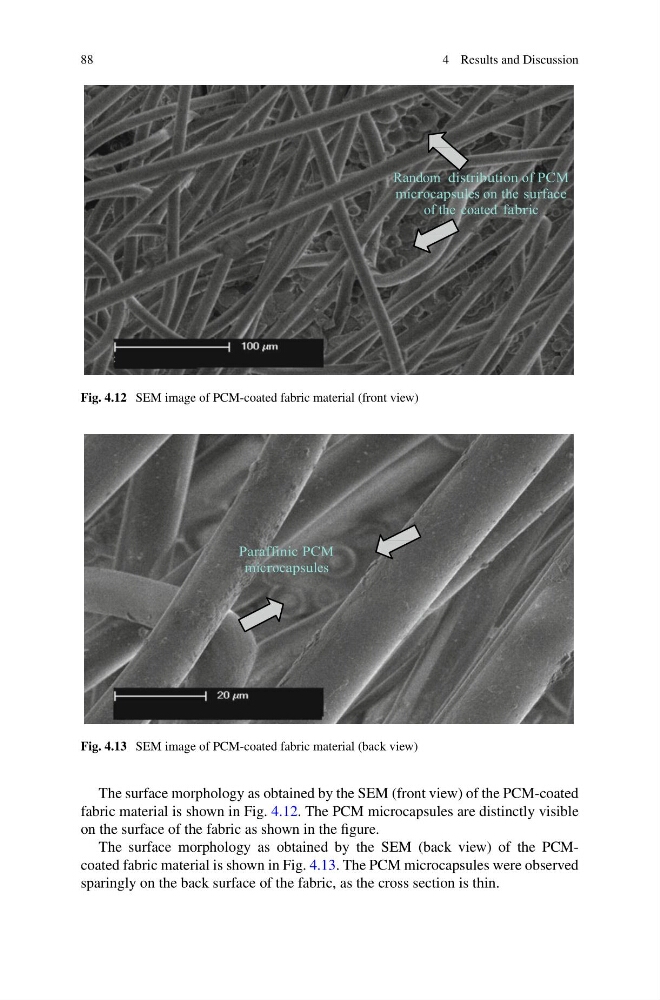
Fig. 4.12 SEM image of PCM-coated fabric material (front view)
Paraffinic PCM
microcapsules
Fig. 4.13 SEM image of PCM-coated fabric material (back view)
The surface morphology as obtained by the SEM (front view) of the PCM-coated
fabric material is shown in Fig. 4.12. The PCM microcapsules are distinctly visible
on the surface of the fabric as shown in the figure.
The surface morphology as obtained by the SEM (back view) of the PCMcoated fabric material is shown in Fig. 4.13. The PCM microcapsules were observed
sparingly on the back surface of the fabric, as the cross section is thin.
4 Results and Discussion
Random distribution of PCM
microcapsules on the surface
of the coated fabric
Fig. 4.12 SEM image of PCM-coated fabric material (front view)
Paraffinic PCM
microcapsules
Fig. 4.13 SEM image of PCM-coated fabric material (back view)
The surface morphology as obtained by the SEM (front view) of the PCM-coated
fabric material is shown in Fig. 4.12. The PCM microcapsules are distinctly visible
on the surface of the fabric as shown in the figure.
The surface morphology as obtained by the SEM (back view) of the PCMcoated fabric material is shown in Fig. 4.13. The PCM microcapsules were observed
sparingly on the back surface of the fabric, as the cross section is thin.