

poorer than 2.5 Å. For example, Paula and Ed solved the structure of 20S proteasome in complex with the inhibitor (EMD-3231) at *3.6 Å resolution [114] as
shown in Fig. 7a. Another example is the structure of 70S ribosome from
Escherichia coli at 2.9 Å resolution in complex with elongation factor Tu,
aminoacyl-tRNA, and the antibiotic kirromycin [115] as shown in Fig. 7b. With
these examples, it is very clear that, in the future, the single-particle cryo-EM will
play a very important role in the preclinical SBDD studies.
PETG
Trp999
Phe601
His418
Glu461
(b)
(a)
Asn102
Met502
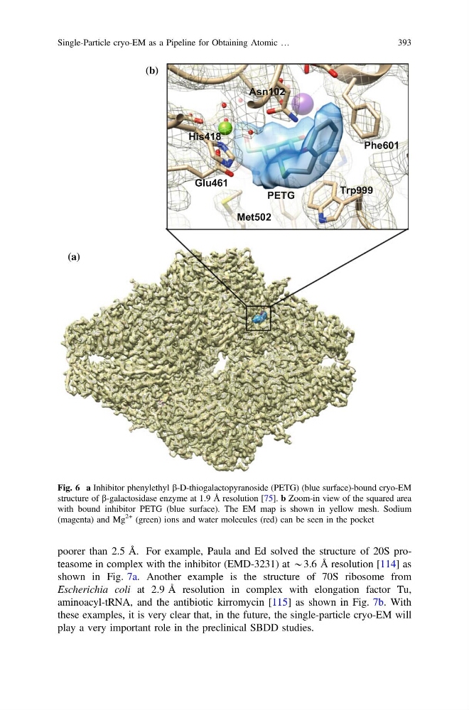
Fig. 6 a Inhibitor phenylethyl b-D-thiogalactopyranoside (PETG) (blue surface)-bound cryo-EM
structure of b-galactosidase enzyme at 1.9 Å resolution [75]. b Zoom-in view of the squared area
with bound inhibitor PETG (blue surface). The EM map is shown in yellow mesh. Sodium
(magenta) and Mg
2+ (green) ions and water molecules (red) can be seen in the pocket
Single-Particle cryo-EM as a Pipeline for Obtaining Atomic …
393
shown in Fig. 7a. Another example is the structure of 70S ribosome from
Escherichia coli at 2.9 Å resolution in complex with elongation factor Tu,
aminoacyl-tRNA, and the antibiotic kirromycin [115] as shown in Fig. 7b. With
these examples, it is very clear that, in the future, the single-particle cryo-EM will
play a very important role in the preclinical SBDD studies.
PETG
Trp999
Phe601
His418
Glu461
(b)
(a)
Asn102
Met502
Fig. 6 a Inhibitor phenylethyl b-D-thiogalactopyranoside (PETG) (blue surface)-bound cryo-EM
structure of b-galactosidase enzyme at 1.9 Å resolution [75]. b Zoom-in view of the squared area
with bound inhibitor PETG (blue surface). The EM map is shown in yellow mesh. Sodium
(magenta) and Mg
2+ (green) ions and water molecules (red) can be seen in the pocket
Single-Particle cryo-EM as a Pipeline for Obtaining Atomic …
393