

Once the grid is on the stage of the TEM, the data is collected on a highly
sensitive direct detection device (DDD) also called as direct electron detector
(DED) under low electron dose (typically <15 e
−
/Å
2 ). Low electron dose is necessary since high dose (>15–20 e
− /Å
2 ) will cause radiation damage. However, high
dose *1000 e
− /Å
2 is required for atomic-resolution reconstruction [25]. This
problem can be overcome by averaging similar looking particles as described in
image processing Sect. 2.3 below. DDD is more sensitive (technically, this feature
is called improved detective quantum efficiency (DQE)) and can detect lower doses
more effectively with low noise as compared to the conventional photographic film
or the CCD (charged coupled device) detectors. Data collection at the focus gives
the best resolution, but however the phase contrast is lost in the image (i.e., you
cannot clearly visualize the particles). In order to visualize the particles, the images
are captured at a defocus that restores the phase contrast in the image, which
enables us to visualize particles. Hence, data is collected at a range of defocus
between *4 µm (lower resolution) and *1 µm (higher resolution). Modern-day
advancements in hardware have led to the use of phase plates and energy filters that
can restore contrast in the images collected closer to focus. Thus, preserving
high-resolution information in the images and at the same time preserving the
image phase/amplitude contract as a result alleviate the need for contrast transfer
function (CTF) modulation correction at image processing stage.
For high-resolution structure determination, the data is collected on DED as
movie frames, which is actually a dose fractionated image stack. The movie frames
collected can be corrected for loss of resolution due to stage drifts, charging, and
beam-induced motion. The individual movie frames or subset of movie frames in
batches are then aligned with respect to each other in order to restore the
high-resolution information [26]. Relatively, high exposures up to 20 e
− /Å
2 can be
used for movie mode while DEDs can also be used in electron counting mode
where dose rate must be kept below 10 e
− /pixel/s [26, 27]. Movie corrections are
applied immediately on the micrographs after the data collection using programs
like MotionCor2 [28], optical flow algorithm as implemented in Xmipp [29, 30],
Unblur/Summovie [31, 32]. In addition, improved stability of specimen can be
provided by the use of grids with graphene and gold support [25, 33, 34]. Hence, in
the last six years there has been many breakthroughs in detector, imaging, and
image processing technology that has led to high-resolution data collection for even
smaller proteins like hemoglobin with mass 64 kDa using Volta phase plate
(VPP) [18], thus leading to resolution revolution with structures determination to
better than 2.5 Å. Another aspect of data collection is the automation. Not all
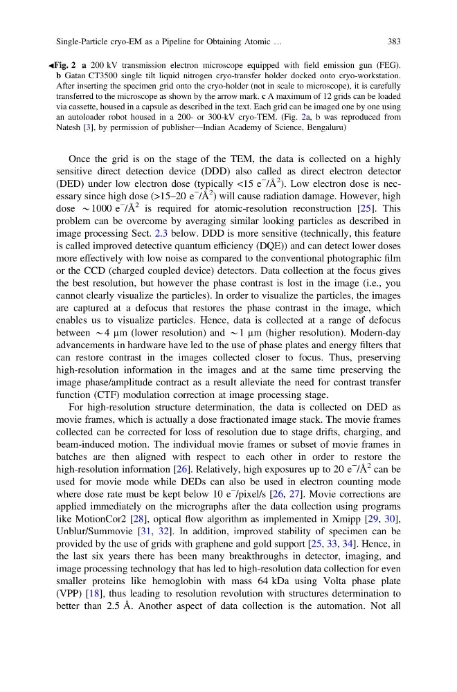
JFig. 2 a 200 kV transmission electron microscope equipped with field emission gun (FEG).
b Gatan CT3500 single tilt liquid nitrogen cryo-transfer holder docked onto cryo-workstation.
After inserting the specimen grid onto the cryo-holder (not in scale to microscope), it is carefully
transferred to the microscope as shown by the arrow mark. c A maximum of 12 grids can be loaded
via cassette, housed in a capsule as described in the text. Each grid can be imaged one by one using
an autoloader robot housed in a 200- or 300-kV cryo-TEM. (Fig. 2a, b was reproduced from
Natesh [3], by permission of publisher—Indian Academy of Science, Bengaluru)
Single-Particle cryo-EM as a Pipeline for Obtaining Atomic …
383
sensitive direct detection device (DDD) also called as direct electron detector
(DED) under low electron dose (typically <15 e
−
/Å
2 ). Low electron dose is necessary since high dose (>15–20 e
− /Å
2 ) will cause radiation damage. However, high
dose *1000 e
− /Å
2 is required for atomic-resolution reconstruction [25]. This
problem can be overcome by averaging similar looking particles as described in
image processing Sect. 2.3 below. DDD is more sensitive (technically, this feature
is called improved detective quantum efficiency (DQE)) and can detect lower doses
more effectively with low noise as compared to the conventional photographic film
or the CCD (charged coupled device) detectors. Data collection at the focus gives
the best resolution, but however the phase contrast is lost in the image (i.e., you
cannot clearly visualize the particles). In order to visualize the particles, the images
are captured at a defocus that restores the phase contrast in the image, which
enables us to visualize particles. Hence, data is collected at a range of defocus
between *4 µm (lower resolution) and *1 µm (higher resolution). Modern-day
advancements in hardware have led to the use of phase plates and energy filters that
can restore contrast in the images collected closer to focus. Thus, preserving
high-resolution information in the images and at the same time preserving the
image phase/amplitude contract as a result alleviate the need for contrast transfer
function (CTF) modulation correction at image processing stage.
For high-resolution structure determination, the data is collected on DED as
movie frames, which is actually a dose fractionated image stack. The movie frames
collected can be corrected for loss of resolution due to stage drifts, charging, and
beam-induced motion. The individual movie frames or subset of movie frames in
batches are then aligned with respect to each other in order to restore the
high-resolution information [26]. Relatively, high exposures up to 20 e
− /Å
2 can be
used for movie mode while DEDs can also be used in electron counting mode
where dose rate must be kept below 10 e
− /pixel/s [26, 27]. Movie corrections are
applied immediately on the micrographs after the data collection using programs
like MotionCor2 [28], optical flow algorithm as implemented in Xmipp [29, 30],
Unblur/Summovie [31, 32]. In addition, improved stability of specimen can be
provided by the use of grids with graphene and gold support [25, 33, 34]. Hence, in
the last six years there has been many breakthroughs in detector, imaging, and
image processing technology that has led to high-resolution data collection for even
smaller proteins like hemoglobin with mass 64 kDa using Volta phase plate
(VPP) [18], thus leading to resolution revolution with structures determination to
better than 2.5 Å. Another aspect of data collection is the automation. Not all
JFig. 2 a 200 kV transmission electron microscope equipped with field emission gun (FEG).
b Gatan CT3500 single tilt liquid nitrogen cryo-transfer holder docked onto cryo-workstation.
After inserting the specimen grid onto the cryo-holder (not in scale to microscope), it is carefully
transferred to the microscope as shown by the arrow mark. c A maximum of 12 grids can be loaded
via cassette, housed in a capsule as described in the text. Each grid can be imaged one by one using
an autoloader robot housed in a 200- or 300-kV cryo-TEM. (Fig. 2a, b was reproduced from
Natesh [3], by permission of publisher—Indian Academy of Science, Bengaluru)
Single-Particle cryo-EM as a Pipeline for Obtaining Atomic …
383