

histones) for transcription factor [37]. Under normal circumstances in plasmodium
parasite, only a few genes are expressed for transcription while the rest remain silent
at a particular stage, and this phenomenon is known as stage-specific expression of
genes. The inhibition of HDAC enzyme increases the acetylation that results in the
loss of control over the gene expression which must be expressed in stage-specific
manner in the parasite and, finally, implode the transcription cascade of parasite
[37]. This enzyme is an attractive target for antimalarial drug therapy because,
unlike mammalian cells, HDACs are more limited and potentially less redundant
than plasmodium species [38].
The crystal structure of PfHDAC-1 is yet to be elucidated. Therefore, a ligandrefined homology model of PfHDAC-1 complexed with a hydroxamate-based
inhibitor TrichostatinA (TSA) was generated, using human HDAC8 as template. It
was observed that the modeled PfHDAC-1 enzyme comprises of a single domain
with open a/b class topology. The structure consists of eight b sheets surrounded by
fourteen a helices and these secondary structures were linked by seven loops.
PfHDAC-1 comprises of hydrophobic upper region (lined with His24, Pro25,
Thr96, Phe148, Phe203, Leu269, and Tyr301 residues) and a Zn
2+ metal ion present in its catalytic site. This metal ion forms penta-coordinated geometry forming
three bonds with the enzyme (side chain O(d) of Asp174, Asp262, and the N(d) of
His176) and two bonds with ligand’s hydroxamate group (i.e., carboxyl and
hydroxyl oxygen) [39].
Mukherjee et al. in 2008 performed molecular docking and molecular dynamics
studies on the homology model and found that hydroxamate group is essential for
binding with PfHDAC enzyme. The carbonyl oxygen of hydroxamate group forms
hydrogen bond interactions with Tyr301, His138, and His139 residues. The TSA
showed hydrophobic interaction with His24, Pro25, Phe148, Phe203, Leu269, and
Tyr301 residues [39].
The development of compounds that are selective for parasitic HDACs over
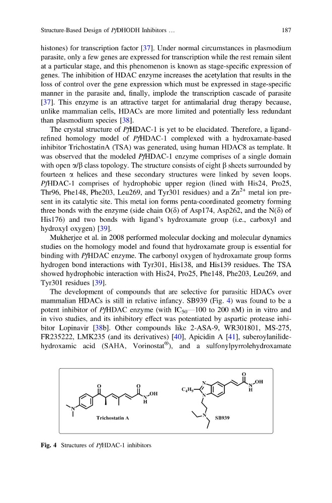
mammalian HDACs is still in relative infancy. SB939 (Fig. 4) was found to be a
potent inhibitor of PfHDAC enzyme (with IC 50 —100 to 200 nM) in in vitro and
in vivo studies, and its inhibitory effect was potentiated by aspartic protease inhibitor Lopinavir [38b]. Other compounds like 2-ASA-9, WR301801, MS-275,
FR235222, LMK235 (and its derivatives) [40], Apicidin A [41], suberoylanilidehydroxamic acid (SAHA, Vorinostat
® ), and a sulfonylpyrrolehydroxamate
Fig. 4 Structures of PfHDAC-1 inhibitors
Structure-Based Design of PfDHODH Inhibitors …
187
parasite, only a few genes are expressed for transcription while the rest remain silent
at a particular stage, and this phenomenon is known as stage-specific expression of
genes. The inhibition of HDAC enzyme increases the acetylation that results in the
loss of control over the gene expression which must be expressed in stage-specific
manner in the parasite and, finally, implode the transcription cascade of parasite
[37]. This enzyme is an attractive target for antimalarial drug therapy because,
unlike mammalian cells, HDACs are more limited and potentially less redundant
than plasmodium species [38].
The crystal structure of PfHDAC-1 is yet to be elucidated. Therefore, a ligandrefined homology model of PfHDAC-1 complexed with a hydroxamate-based
inhibitor TrichostatinA (TSA) was generated, using human HDAC8 as template. It
was observed that the modeled PfHDAC-1 enzyme comprises of a single domain
with open a/b class topology. The structure consists of eight b sheets surrounded by
fourteen a helices and these secondary structures were linked by seven loops.
PfHDAC-1 comprises of hydrophobic upper region (lined with His24, Pro25,
Thr96, Phe148, Phe203, Leu269, and Tyr301 residues) and a Zn
2+ metal ion present in its catalytic site. This metal ion forms penta-coordinated geometry forming
three bonds with the enzyme (side chain O(d) of Asp174, Asp262, and the N(d) of
His176) and two bonds with ligand’s hydroxamate group (i.e., carboxyl and
hydroxyl oxygen) [39].
Mukherjee et al. in 2008 performed molecular docking and molecular dynamics
studies on the homology model and found that hydroxamate group is essential for
binding with PfHDAC enzyme. The carbonyl oxygen of hydroxamate group forms
hydrogen bond interactions with Tyr301, His138, and His139 residues. The TSA
showed hydrophobic interaction with His24, Pro25, Phe148, Phe203, Leu269, and
Tyr301 residues [39].
The development of compounds that are selective for parasitic HDACs over
mammalian HDACs is still in relative infancy. SB939 (Fig. 4) was found to be a
potent inhibitor of PfHDAC enzyme (with IC 50 —100 to 200 nM) in in vitro and
in vivo studies, and its inhibitory effect was potentiated by aspartic protease inhibitor Lopinavir [38b]. Other compounds like 2-ASA-9, WR301801, MS-275,
FR235222, LMK235 (and its derivatives) [40], Apicidin A [41], suberoylanilidehydroxamic acid (SAHA, Vorinostat
® ), and a sulfonylpyrrolehydroxamate
Fig. 4 Structures of PfHDAC-1 inhibitors
Structure-Based Design of PfDHODH Inhibitors …
187