

minimization and finally experimental determination of binding affinity and crystallization of complex structure was used. This iterative process yielded a series of
potent and membrane-permeable 9-(arylmethyl)-9-deazapurines (2-amino-7-(arylmethyl)-4H-pyrrolo[3,2-d]-pyrimidin-4-ones) inhibitors of PNP [29]. Later, (S)-9[l-(3-chlorophenyl)-2-carboxyethyl]-9-deazaguanin showed highest potency among
all previously designed analogs [32]; however, the (R)-isomer was 30-fold less
potent. This study exemplifies how structural information can be carefully used
toward designing of potent inhibitors of the receptor of interest.
The enthalpy and entropy components of binding free energy together decide
affinity of interaction between receptor and ligand. Therefore, affinity can be modulated favorably adopting following possible strategies: (i) decreasing the unfavorable
entropy maintaining favorable enthalpy, (ii) increasing favorable enthalpy without
introducing unfavorable entropy, and (iii) altering one or both of enthalpy and entropy
favorably without losing proportionally on other component [33].
An example where first strategy has been used for optimizing affinity is inhibitors of PNP. Optimized picomolar-binding PNP inhibitors have also been
reported [34]. The attention has been paid on reducing the entropic penalty, without
sacrificing the enthalpy of binding to gain affinity. Hypoxanthine has K i 4.3 lM,
with enthalpy −30.5 kcal/mol, but 23.1 kcal/mol entropy penalty to result a
−7.4 kcal/mol binding free energy [35], but optimized molecule SerMe-ImmH
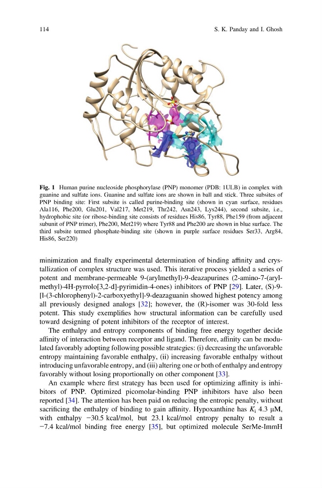
Fig. 1 Human purine nucleoside phosphorylase (PNP) monomer (PDB: 1ULB) in complex with
guanine and sulfate ions. Guanine and sulfate ions are shown in ball and stick. Three subsites of
PNP binding site: First subsite is called purine-binding site (shown in cyan surface, residues
Ala116, Phe200, Glu201, Val217, Met219, Thr242, Asn243, Lys244), second subsite, i.e.,
hydrophobic site (or ribose-binding site consists of residues His86, Tyr88, Phe159 (from adjacent
subunit of PNP trimer), Phe200, Met219) where Tyr88 and Phe200 are shown in blue surface. The
third subsite termed phosphate-binding site (shown in purple surface residues Ser33, Arg84,
His86, Ser220)
114
S. K. Panday and I. Ghosh
potent and membrane-permeable 9-(arylmethyl)-9-deazapurines (2-amino-7-(arylmethyl)-4H-pyrrolo[3,2-d]-pyrimidin-4-ones) inhibitors of PNP [29]. Later, (S)-9[l-(3-chlorophenyl)-2-carboxyethyl]-9-deazaguanin showed highest potency among
all previously designed analogs [32]; however, the (R)-isomer was 30-fold less
potent. This study exemplifies how structural information can be carefully used
toward designing of potent inhibitors of the receptor of interest.
The enthalpy and entropy components of binding free energy together decide
affinity of interaction between receptor and ligand. Therefore, affinity can be modulated favorably adopting following possible strategies: (i) decreasing the unfavorable
entropy maintaining favorable enthalpy, (ii) increasing favorable enthalpy without
introducing unfavorable entropy, and (iii) altering one or both of enthalpy and entropy
favorably without losing proportionally on other component [33].
An example where first strategy has been used for optimizing affinity is inhibitors of PNP. Optimized picomolar-binding PNP inhibitors have also been
reported [34]. The attention has been paid on reducing the entropic penalty, without
sacrificing the enthalpy of binding to gain affinity. Hypoxanthine has K i 4.3 lM,
with enthalpy −30.5 kcal/mol, but 23.1 kcal/mol entropy penalty to result a
−7.4 kcal/mol binding free energy [35], but optimized molecule SerMe-ImmH
Fig. 1 Human purine nucleoside phosphorylase (PNP) monomer (PDB: 1ULB) in complex with
guanine and sulfate ions. Guanine and sulfate ions are shown in ball and stick. Three subsites of
PNP binding site: First subsite is called purine-binding site (shown in cyan surface, residues
Ala116, Phe200, Glu201, Val217, Met219, Thr242, Asn243, Lys244), second subsite, i.e.,
hydrophobic site (or ribose-binding site consists of residues His86, Tyr88, Phe159 (from adjacent
subunit of PNP trimer), Phe200, Met219) where Tyr88 and Phe200 are shown in blue surface. The
third subsite termed phosphate-binding site (shown in purple surface residues Ser33, Arg84,
His86, Ser220)
114
S. K. Panday and I. Ghosh