

198
S. Sakkiah et al.
and dynamics simulation techniques elucidated binding interactions between the
human α4β2 and tobacco constituents [68].
Initially, the 3D structure of the human α4β2 extracellular domain was constructed
utilizing the crystal structure of Ct-AChBP and homology modeling. MD simulations
were used to minimize the constructed human α4β2 3D structure. Eleven components which were both crystallized with nAChRs or AChBP and also have binding
data for α4β2 were selected to dock with α4β2 protein. The LigPrep2.0 tool from
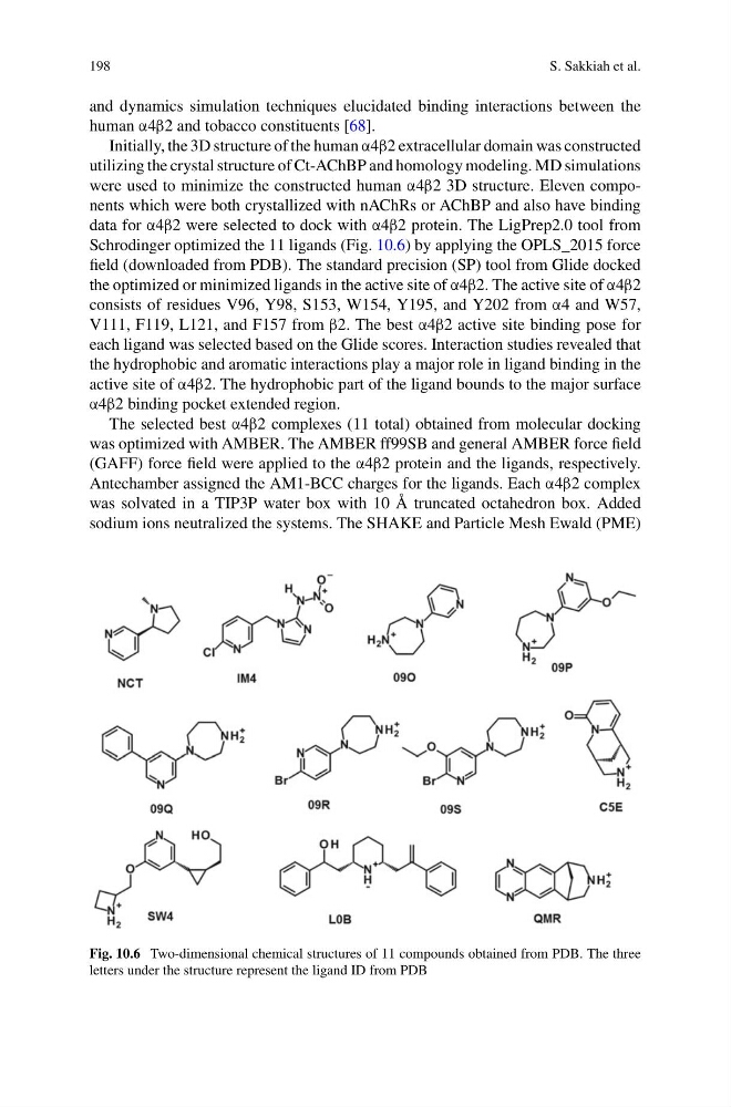
Schrodinger optimized the 11 ligands (Fig. 10.6) by applying the OPLS_2015 force
field (downloaded from PDB). The standard precision (SP) tool from Glide docked
the optimized or minimized ligands in the active site of α4β2. The active site of α4β2
consists of residues V96, Y98, S153, W154, Y195, and Y202 from α4 and W57,
V111, F119, L121, and F157 from β2. The best α4β2 active site binding pose for
each ligand was selected based on the Glide scores. Interaction studies revealed that
the hydrophobic and aromatic interactions play a major role in ligand binding in the
active site of α4β2. The hydrophobic part of the ligand bounds to the major surface
α4β2 binding pocket extended region.
The selected best α4β2 complexes (11 total) obtained from molecular docking
was optimized with AMBER. The AMBER ff99SB and general AMBER force field
(GAFF) force field were applied to the α4β2 protein and the ligands, respectively.
Antechamber assigned the AM1-BCC charges for the ligands. Each α4β2 complex
was solvated in a TIP3P water box with 10 Å truncated octahedron box. Added
sodium ions neutralized the systems. The SHAKE and Particle Mesh Ewald (PME)
Fig. 10.6 Two-dimensional chemical structures of 11 compounds obtained from PDB. The three
letters under the structure represent the ligand ID from PDB
S. Sakkiah et al.
and dynamics simulation techniques elucidated binding interactions between the
human α4β2 and tobacco constituents [68].
Initially, the 3D structure of the human α4β2 extracellular domain was constructed
utilizing the crystal structure of Ct-AChBP and homology modeling. MD simulations
were used to minimize the constructed human α4β2 3D structure. Eleven components which were both crystallized with nAChRs or AChBP and also have binding
data for α4β2 were selected to dock with α4β2 protein. The LigPrep2.0 tool from
Schrodinger optimized the 11 ligands (Fig. 10.6) by applying the OPLS_2015 force
field (downloaded from PDB). The standard precision (SP) tool from Glide docked
the optimized or minimized ligands in the active site of α4β2. The active site of α4β2
consists of residues V96, Y98, S153, W154, Y195, and Y202 from α4 and W57,
V111, F119, L121, and F157 from β2. The best α4β2 active site binding pose for
each ligand was selected based on the Glide scores. Interaction studies revealed that
the hydrophobic and aromatic interactions play a major role in ligand binding in the
active site of α4β2. The hydrophobic part of the ligand bounds to the major surface
α4β2 binding pocket extended region.
The selected best α4β2 complexes (11 total) obtained from molecular docking
was optimized with AMBER. The AMBER ff99SB and general AMBER force field
(GAFF) force field were applied to the α4β2 protein and the ligands, respectively.
Antechamber assigned the AM1-BCC charges for the ligands. Each α4β2 complex
was solvated in a TIP3P water box with 10 Å truncated octahedron box. Added
sodium ions neutralized the systems. The SHAKE and Particle Mesh Ewald (PME)
Fig. 10.6 Two-dimensional chemical structures of 11 compounds obtained from PDB. The three
letters under the structure represent the ligand ID from PDB